also: cerebellar cortex
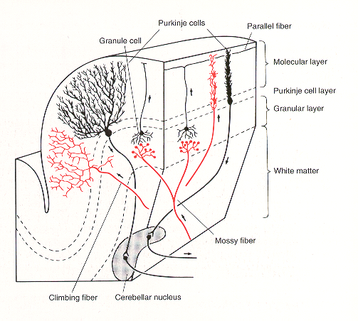
the cerebellar cortex is organized into three layers: the molecular layer, the Purkinje cell layer, and the granule layer. The cerebellar cortex contains different types of neurons, including stellate neurons, basket neurons, Purkinje neurons, Golgi neurons, and granule cells. Granule cells are the only excitatory neurons in the cerebellar cortex. The cerebellar cortex is folded and has many parallel convolutions called folia.
- 10% of total brain mass
- 50% of all neurons in the brain
- most of the neurons are granule cells
- no recurrent activation
- largest neuron: inhibitory Purkinje Cell
- The white matter structures are also known as arbor vitae (tree of life).
- the organization in the cerebellum is other than the cerebrum, ipsilateral
What is the Cerebellum for?
- makes sure skilled movements are executed properly and adjusted whenever the execution fails to meet the expectations ⇒ Prediction Error
Cerebellum Circuit
Purkinje cells are oriented in parallel
The mossy fibre / granule cell / parallel fibre system and the dendritic trees of Purkinje cells are arranged orthogonal. Each parallel fibre runs for several millimetres orthogonal through hundreds of nearly 2D Purkinje cells dendrites. In the panel a tangential section is shown with very many parallel fibres (thin lines) crossing orthogonal the Purkinje cell dendritic trees (thick ).
In this way each granule cells / parallel fibre makes few (or a single) contact with very many Purkinje cells.
Climbing fibres, in contrast, contact few (10) Purkinje cells with many synapses.
- umschlingt den Dendritenbaum einer Purkinje-Zelle komplett
- bildet eine 1:1 Verbindung zu genau EINER Purkinje-Zelle
- macht 300–600 synaptische Kontakte mit dieser einen Purkinje-Zelle
Lobes
- Anterior Lobe
- Posterior Lobe
- Flocculonodular lobe
Lesion
- Delayed Movement Initiation
- Asking subject with unilateral lesion of the cerebellum to clench both hands results in delayed initiation.
- Dysmetry
- loss of precision of movements, undershoot, overshoot
- Decomposition
- Coordination of multi joint movements is lost, e.g. shoulder moves before elbow, sequential instead of joint movements
- Intentional Tremor
- jittering movements get stronger when target is approached.
- Dysdiadochokinesia
- inability to perform rapidly alternating antagonistic movements, e.g. pronation and supination of the lower arm.
- Hypotonia
- reduction of resistance on passive limb movements (low muscle tone)
- Nystagmus
- jittering eye movements
- Deficits in motor learning
- deficits in movement coordination & classical conditioning
Learning
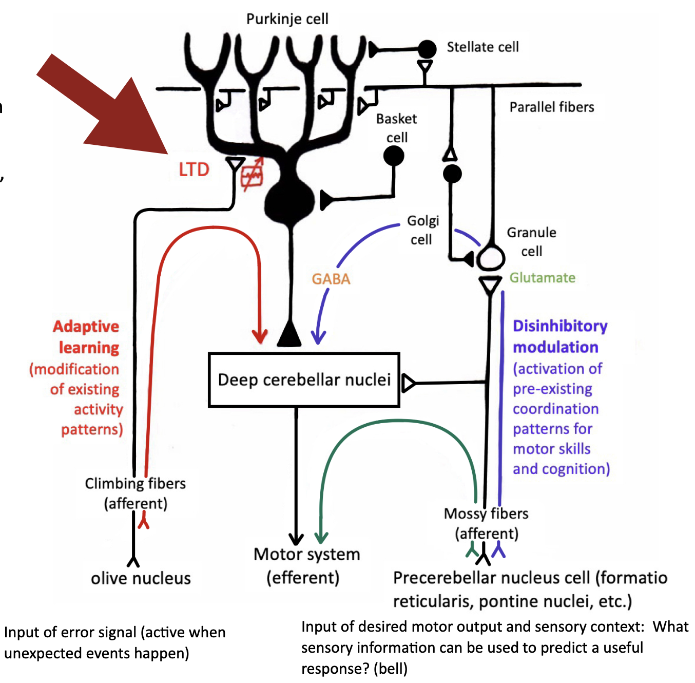
- Learning is gated by the climbing fibres. The synapses of the parallel fibres targeting the Purkinje cells are modified.
- Mossy fibre spontaneous activity leads to steady input, the frequency can be very high and modulated by sensory input. Detailed information can be coded.
- Climbing fibre spontaneous activity has low rates, with only small changes through sensory input. They can be used, however, to code timing of events or work as triggers.
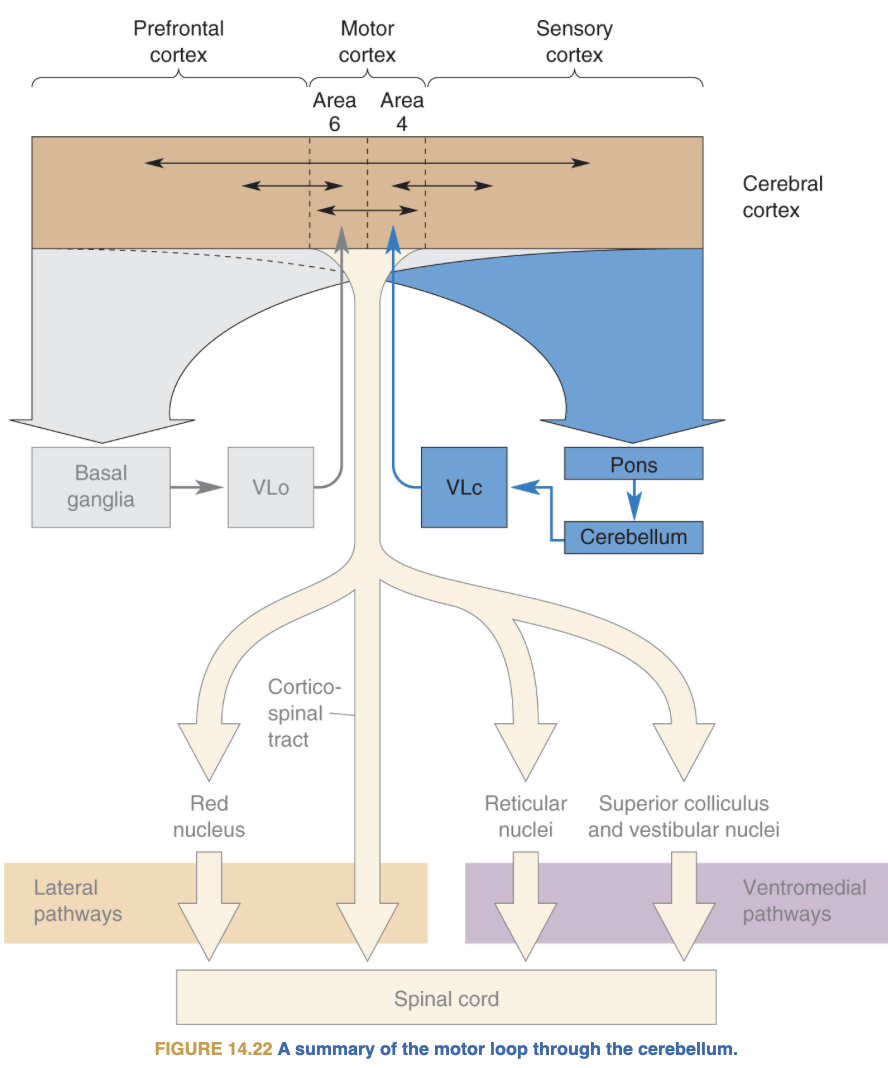
Connections to non-motor areas
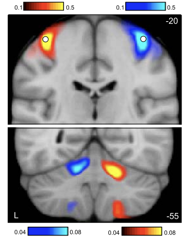
The cerebellum is functionally connected to non-motor areas (Prefrontal-, Posterior-Parietal-Cortex, basal ganglia) and is activated in non-motor tasks.
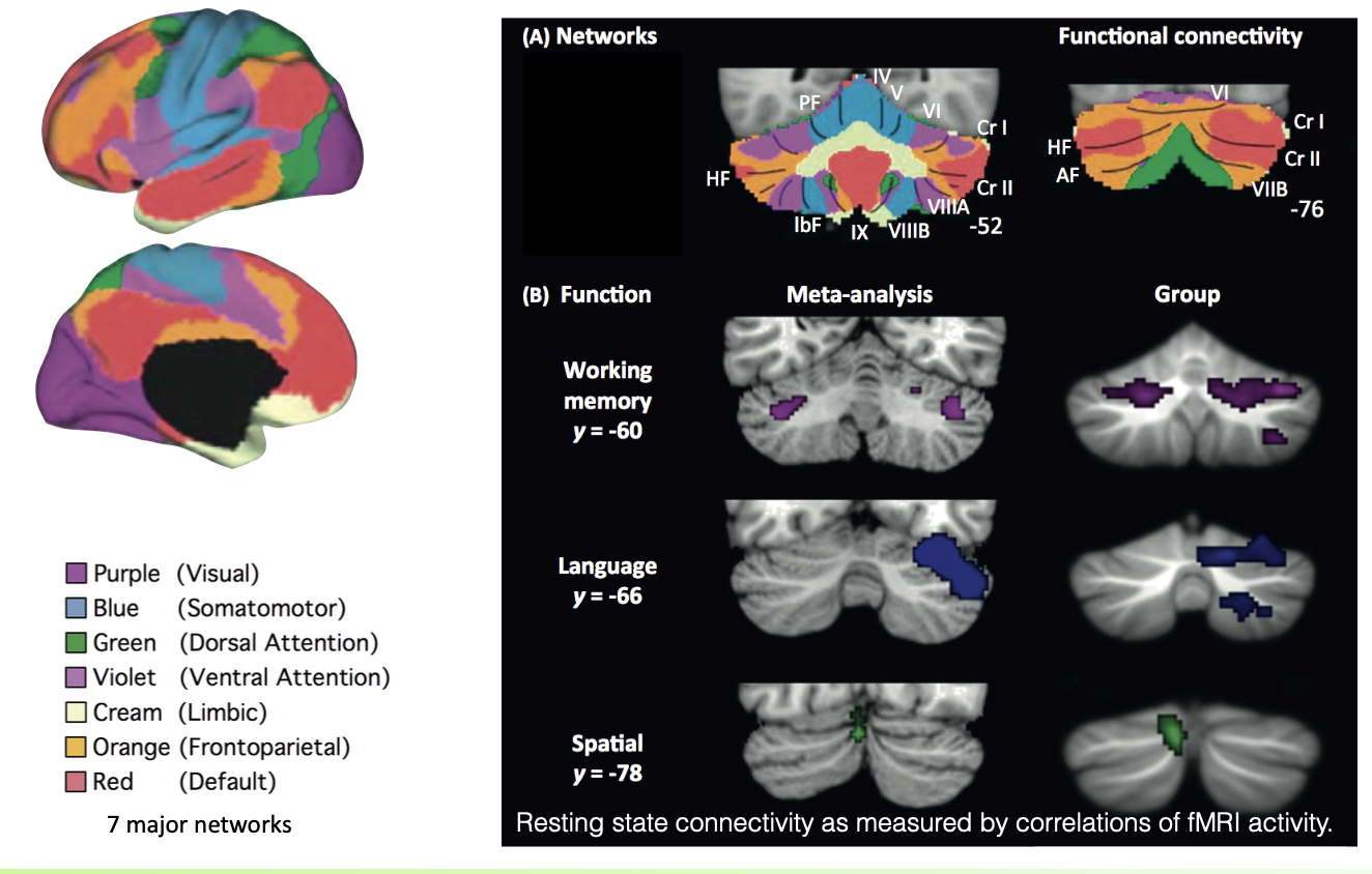
New view: widespread involvement in non-motor function
- Lesions lead to motor deficits, but also to deficits in emotion, working memory, language, divided attention
- Functional Imaging Studies reveal non-motor-related activation in the Cerebellum
- Anatomical studies reveal a separation of the dentate nucleus, in motor-and non-motor related connections and connections to PFC, Posterior Parietal Cortex, and Basal Ganglia.
Classical Conditioning in Cerebellum
- the neutral/conditioned stimulus (CS) gets into the cerebellum via the mossy fibers (from cortex, spinal cord, vestibular system)
- the unconditioned stimulus (US) comes in via the climbing fibers (from the Inferior Olive), changing the synapses of the parallel fibers targeting the Purkinje Cells.
- The activity patterns across granule cells serves as a clock and the model reproduces the appropriate timing of the conditioned response.
- The conditioned response builds up over a few training sessions, with visible training effect of both nucleus and Purkinje cell activity.
Differences to Cerebrum
Cerebrum:
- contralateral organisation
- 20% inhibitory neurons
- Excitatory input and output
- Most areas (V1, V2, FFA, PPA,…) have excitatory connections with each other
- The backbone of recurrent excitation
- The cerebrum is ‘mostly talking to itself.’
Cerebellum: - Excitatory input, inhibitory output (Purkinje cells onto the deep cerebellar nuclei)
- No recurrent activation
- Request from outside, computation, output
- No self-sustained activation over longer periods of time (on-demand computations)
- Without external input, there is no activity. This is in marked contrast to the cerebrum
Why is the cerebellum a feed forward system?
Transclude of Why-is-the-cerebellum-a-feed-forward-system
see also
Tags: neuroscience science gehirnregion
Superlink: 050 🧠Neuroscience
Parietal lobe
Mirror Neurons (MN)
Quellen
Erstellt: 25-02-22 17:51