Neurotransmission
also see Synaptic Transmission
Overview of Neurotransmission
chatbot
Neurotransmission is the process by which nerve cells, or neurons, communicate with each other or with target cells such as muscle fibers. This communication occurs at specialized junctions called synapses and involves both electrical and chemical signals.
Types of Synapses
-
Chemical Synapses (Electrochemical Synapses):
- Mechanism:
- An action potential reaches the presynaptic terminal, causing voltage-gated calcium channels to open.
- Calcium ions enter the presynaptic terminal, triggering the fusion of synaptic vesicles with the presynaptic membrane.
- Neurotransmitters are released into the synaptic cleft and bind to receptors on the postsynaptic membrane, altering its membrane potential.
- Signal transmission is terminated by the degradation or reuptake of neurotransmitters.
- Advantages:
- Allows for amplification, modulation, and diversity in signal transmission.
- Supports synaptic plasticity and unidirectional transmission.
- Disadvantages:
- Slower transmission due to multiple steps.
- Requires energy for maintaining gradients and neurotransmitter release.
- Mechanism:
-
Electrical Synapses:
- Mechanism:
- Direct electrical coupling between cells via gap junctions composed of connexons.
- Allows ions and small molecules to flow directly between cells.
- Advantages:
- Very fast transmission and bidirectional signaling.
- Facilitates synchronization of neuronal networks.
- Low energy requirement.
- Disadvantages:
- Limited amplification and modulation.
- Less flexibility in information processing.
- Mechanism:
Key Components in Chemical Synapses
-
Synaptic Vesicles:
- Contain neurotransmitters such as acetylcholine (ACh).
- Store and release neurotransmitters into the synaptic cleft upon stimulation.
-
Neurotransmitter Release:
- Triggered by the influx of calcium ions following an action potential.
- Involves the fusion of synaptic vesicles with the presynaptic membrane (exocytosis).
-
Synaptic Cleft and Receptors:
- Neurotransmitters diffuse across the synaptic cleft and bind to specific receptors on the postsynaptic membrane.
- Receptors can be ion channels or G-protein-coupled receptors.
-
Endplate Potential:
- The binding of neurotransmitters induces a small membrane depolarization known as the endplate potential.
- Can trigger a muscle action potential through voltage-gated sodium channels.
-
Termination of Signal:
- Achieved by enzymatic degradation of neurotransmitters, such as acetylcholine by acetylcholine-esterase (AChE).
cycle.png)
- Achieved by enzymatic degradation of neurotransmitters, such as acetylcholine by acetylcholine-esterase (AChE).
Proteins Involved in Neurotransmission
-
Synapsin:
- Anchors synaptic vesicles to microfilaments in the resting state.
- Releases vesicles upon calcium influx, preparing them for fusion.
-
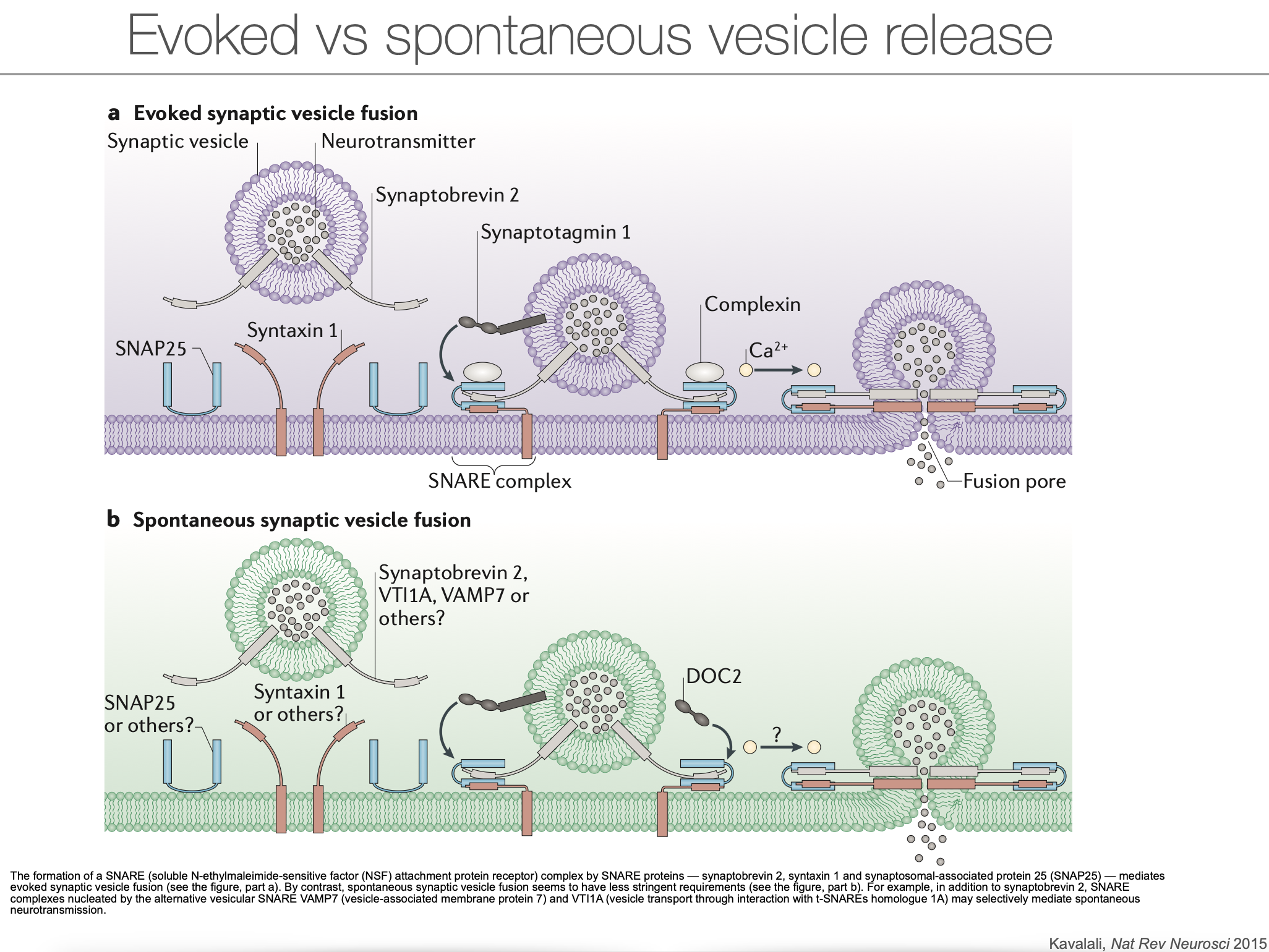
SNARE Proteins:
- Synaptobrevin (v-SNARE): Embedded in vesicle membranes, involved in membrane fusion.
- Syntaxin (t-SNARE): Located in the presynaptic membrane, forms the fusion complex with v-SNAREs.
- Synaptotagmin: Acts as a calcium sensor, initiating vesicle fusion upon calcium binding.
Summary
Neurotransmission is a complex process involving the precise coordination of electrical and chemical signals. Chemical synapses allow for diverse and modifiable communication, while electrical synapses provide rapid and synchronized signaling. Understanding these mechanisms is crucial for insights into neural communication and potential therapeutic targets for neurological disorders.
Sources:
- Intro to Neurobiology Script
- Synaptic Transmission
- Axon (Nerve) Terminal
- Neurotransmission
- Synapse
- Action Potential
![]()
- nerve stimulation cause a rise of intracellular Ca2+ concentration at the presynaptic terminal (calcium sensors);
- neurotransmitter release can be induced in the absence of action potentials or Ca2+ entry from the extracellular media (Ca2+ caging compounds - light stimulation)
- single action potentials generate a Ca2+ rise as little as 10 nM, which lasts up to a few seconds. This increment in [Ca2+] is a small fraction of the typical resting [Ca2+] of 100 nM;
- given the short latency between Ca2+ entry and postsynaptic events, there must be a pool of synaptic vesicles that are ready to fuse with the presynaptic plasma membrane immediately upon a rise of intracellular Ca2+ concentration.
Neurotransmitter release in numbers
- The fusion of one vesicle releases about 5000 neurotransmitter molecules within a millisecond that produce a quantal response in the postsynaptic site.
- At neuromuscular junctions, transmitter from one vesicle diffuses across the synaptic cleft in 2μs and reaches a concentration of about 1mM at the postsynaptic receptors.
- These receptors bind transmitter rapidly, opening from 1000 to 2000 postsynaptic ion channels.
- Each channel has a 25-pS conductance and remains open for about 1.5 ms, admitting a net inflow of 35,000 positive ions.
- A single action potential in a motor neuron can release 300 quanta within about 1.5 ms along a junction that contains about 1000 active zones. The resulting postsynaptic depolarization, which begins after a synaptic delay of about 0.5 ms and reaches a peak of tens of millivolts, is typically sufficient to generate an action potential in the muscle fiber.
- At fast central synapses each action potential releases from 5 to 10 quanta, and each quantum released elevates the transmitter concentration in the cleft to about 1 mM and activates about 30 ion channels.
- At excitatory synapses, this release may be sufficient to generate EPSPs of 1mV or less in amplitude, clearly subthreshold for generating action potentials.
Source: Byrne et al., From molecules to networks, Academic Press
see also
Tags: neuroscience science
Superlink: 050 🧠Neuroscience 051 ☣Neurobiology
Source
Created: 2025-06-11 18:00