Synaptic Transmission
also see Neurotransmission
#chatbot
Synapsin, synaptobrevin, syntaxin, and synaptotagmin are key proteins in neurotransmission, working together in the presynaptic nerve terminal to enable the release of neurotransmitters. They perform distinct yet interrelated functions:
![]()
![]()
-
Synapsin:
- Synapsin is not a SNARE protein; rather, it is a regulatory protein that anchors synaptic **vesicles to microfilaments within the Axon (Nerve) Terminal.
- It secures the vesicles to the microfilaments during the resting phase.
- When the intracellular calcium concentration rises, synapsin changes its conformation, allowing the vesicles to detach from the microfilaments. This prepares the vesicles for subsequent fusion with the membrane.
-
Synaptobrevin:
- Synaptobrevin is a v-SNARE (vesicle-SNARE) protein embedded in the membrane of synaptic vesicles.
- It features an alpha-helix that traverses the vesicle membrane and a globular unit with beta-sheet structures.
- Synaptobrevin binds to t-SNAREs and is directly involved in the fusion of the vesicle with the plasma membrane. Tetanus toxin can cleave synaptobrevin, thereby blocking neurotransmission.
-
Syntaxin:
- Syntaxin is a t-SNARE (target-SNARE) protein located in the presynaptic plasma membrane.
- It has a long alpha-helix structure.
- Syntaxin binds to v-SNAREs like synaptobrevin to form the fusion complex.
-
Synaptotagmin:
- Synaptotagmin is neither a v-SNARE nor a t-SNARE; it is a presynaptic membrane protein that functions as a calcium sensor.
- It has a long alpha-helix structure.
- When the intracellular calcium concentration increases, synaptotagmin binds to calcium, leading to the fusion of vesicles with the plasma membrane.
- It is essential for the formation of the fusion complex.
Connection and Interaction:
- Vesicle Attachment: Synapsin keeps synaptic vesicles attached to microfilaments during the resting phase.
- Calcium Signal: When an action potential reaches the nerve terminal, calcium ions flow into the cell.
- Vesicle Release: The rise in calcium concentration causes a conformational change in synapsin, releasing the vesicles from the microfilaments.
- Formation of the Fusion Complex: v-SNAREs (synaptobrevin) on the vesicles connect with t-SNAREs (syntaxin) on the presynaptic membrane. Synaptotagmin acts as a calcium sensor and, upon binding calcium, initiates fusion.
- Membrane Fusion: The coiling of the alpha-helices of v- and t-SNAREs pulls the vesicle and plasma membranes together, resulting in membrane fusion.
- Neurotransmitter Release: Fusion of the membranes releases neurotransmitters into the synaptic cleft.
In summary, synapsin holds the vesicles ready for release, synaptobrevin (v-SNARE) and syntaxin (t-SNARE) are responsible for actual membrane fusion, and synaptotagmin, acting as a calcium sensor, initiates the fusion process. These proteins work precisely together to ensure the timely and spatially coordinated release of neurotransmitters.
⚡ Three Modes of Synaptic Transmission: A Comparative Framework
| Feature | Electrical Synapse | Ionotropic Synapse | Metabotropic Synapse |
|---|---|---|---|
| Signal Type | Ionic current via gap junctions | Ligand-gated ion channel | G-protein-coupled receptor signaling |
| Speed | Very fast (sub-millisecond) | Fast (1–5 ms) | Slow (hundreds of ms to seconds) |
| Directionality | Often bidirectional | Unidirectional (pre → post) | Unidirectional |
| Synaptic Delay | ~0 ms | ~1–2 ms | ≥100 ms |
| Modifiability | Limited (but some plasticity exists) | Modifiable via receptor number, subunit comp. | Highly modifiable via second messengers |
| Mechanism | Gap junction channels (connexins) | Neurotransmitter opens ion channels | Neurotransmitter activates intracellular cascades |
| Primary Function | Synchronization, fast escape responses | Fast excitation/inhibition | Modulation, amplification, long-term effects |
| Found in | Invertebrates, brainstem, retina | Widespread across CNS and PNS | CNS, modulatory systems (e.g. dopamine, serotonin) |
🧠 Where Do Electrical Synapses Fit?
🔌 They are
not mediated by receptors
.
-
No neurotransmitter is released.
-
No ligand binding occurs.
-
Thus, electrical synapses are fundamentally distinct from both ionotropic and metabotropic mechanisms.
🔗 Instead, they use
gap junctions
, formed by
connexin proteins
, which:
-
Connect the cytoplasm of two cells directly.
-
Allow ions and small molecules (e.g., ATP) to flow passively.
-
Enable rapid synchronization of neural ensembles (e.g., in oscillatory networks, some retinal and brainstem circuits).
🧩 Functional Role in Neural Systems
-
Electrical synapses are especially important for:
-
Synchronous firing (e.g., inhibitory interneurons in cortex or cerebellum).
-
Developmental signaling (e.g., synchronizing immature networks).
-
Rapid reflex circuits (e.g., Mauthner cells in fish escape responses).
-
🧬 Integration with Chemical Synapses
-
Some neurons use both electrical and chemical synapses to fine-tune timing and reliability.
-
Electrical synapses can prime a neuron for faster response to a subsequent ionotropic input.
🔄 Summary
Electrical synapses:
-
Operate outside the receptor paradigm (no ionotropic or metabotropic involvement).
-
Provide direct ionic continuity, unlike ligand-triggered chemical signaling.
-
Serve as the fastest and most reliable form of communication, at the cost of flexibility.
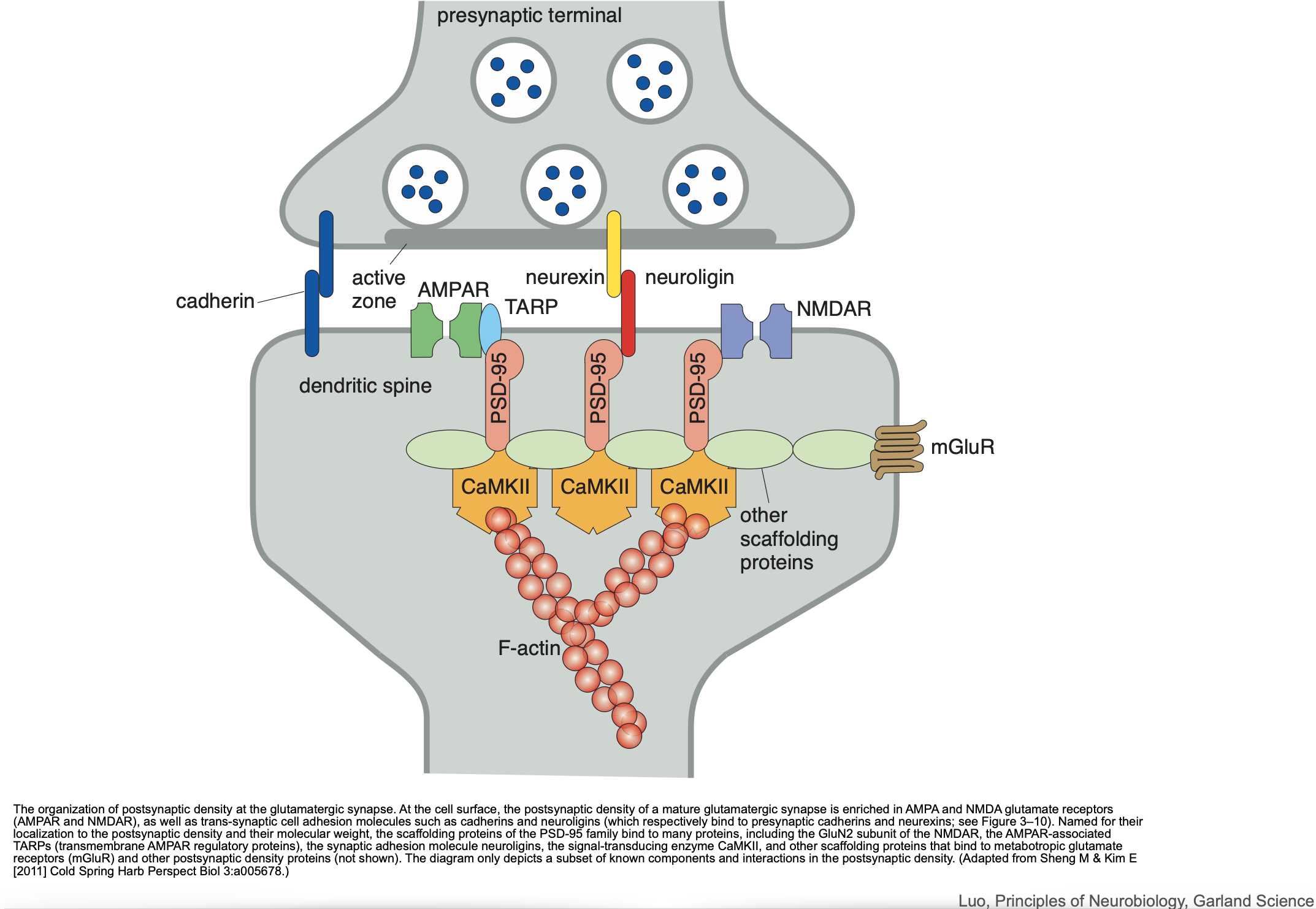
Postsynaptic
see also
Tags: neurobiology science
Superlink: 051 ☣Neurobiology 050 🧠Neuroscience
axo-axonic presynaptic transmission
Source
Created: 12-02-25 12:58