Cellular and Molecular Neuroscience
Summary Lab Report
Paper Discussions CNM
Lectures
Cellular and Molecular Neuroscience – Introduction (01_Introduction.pdf)
Course Objectives
The course is designed to:
-
Deepen understanding of the molecular basis of neuronal function: Students will learn how the intricate interactions among DNA, RNA, proteins, and lipids underlie neural cell function, including processes like synaptic transmission, receptor signaling, and gene expression in neurons.
-
Train students to critically evaluate primary literature: A core skill in neuroscience is the ability to assess experimental design, data interpretation, and theoretical implications in research papers.
-
Connect molecular-level phenomena to disease and therapy: By studying molecular dysfunctions, students will understand how aberrations in gene expression, receptor activity, or protein synthesis contribute to neurological disorders and how they can be targeted pharmacologically.
What is Cellular and Molecular Neuroscience?
Definition and Scope
Cellular and Molecular Neuroscience is a multidisciplinary field focused on:
-
Understanding how neuronal and glial cells function at the molecular level, using tools from molecular biology, biochemistry, and cell biology.
-
Exploring how neurons communicate, grow, differentiate, and form functional networks.
-
Investigating the pathogenesis of neurological and psychiatric disorders through molecular mechanisms such as aberrant protein expression or synaptic dysfunction.
-
Establishing a mechanistic foundation for understanding complex behaviors and mental illness by linking molecular changes to circuit- and behavior-level consequences.
Institutional and Research Relevance
-
University programs (e.g., UCSB) and NIMH branches support integrative research that links cellular and molecular neuroscience to translational and clinical applications.
-
Emphasis is placed on developing mechanistic models of behavior that stem from molecular function, particularly those relevant to psychiatric disease models.
Course Content Overview
Major Thematic Areas
-
Molecular and Cell Biology Foundations: Key concepts include gene structure, the central dogma, molecular interactions, and the chemical logic of cellular function.
-
Neuroscience Techniques: Molecular cloning, electrophoresis, microscopy, CRISPR-Cas9, transgenics, and other methods used to investigate neural structure and function.
-
Cellular Architecture of the Nervous System: Classification and function of neurons and glia, as well as structural components like dendrites, axons, synapses, and myelin.
-
Electrophysiological Principles: Ion gradients, membrane potentials, and the mechanisms underlying neuronal excitability and signal propagation.
-
Synaptic Transmission and Plasticity: Mechanisms of neurotransmitter release, receptor function, and activity-dependent plasticity such as long-term potentiation (LTP).
-
Neurodevelopment: Molecular regulation of neuronal migration, axon guidance, and synaptic refinement.
-
Behavioral Neurogenetics: How molecular pathways influence social behaviors, stress responses, and learning.
Molecular and Cell Biology Concepts
Molecular Biology: Foundational Principles
-
Molecular biology seeks to uncover the molecular logic underlying biological processes. It focuses on:
-
The structure and function of macromolecules such as DNA, RNA, and proteins.
-
The mechanisms by which these molecules interact to execute cellular functions.
-
The molecular basis of regulation, such as transcriptional control, signaling cascades, and protein-protein interactions.
-
-
As William Astbury emphasized, molecular biology is not defined by a specific method but by an approach that looks beneath organismal and cellular phenomena to reveal the molecular ‘blueprint’.
Cell Biology: Organization and Function
-
Cell biology complements molecular biology by focusing on the spatial and structural context in which molecular mechanisms occur.
-
The cell is the basic structural and functional unit of all living organisms.
-
Central themes include:
-
Intracellular compartmentalization (e.g., nucleus, mitochondria, ER)
-
Membrane dynamics and transport
-
Cell signaling and communication
-
Cellular differentiation and specialized function in tissues such as brain
-
Diversity of Cellular Life
Examples of Cellular Variety
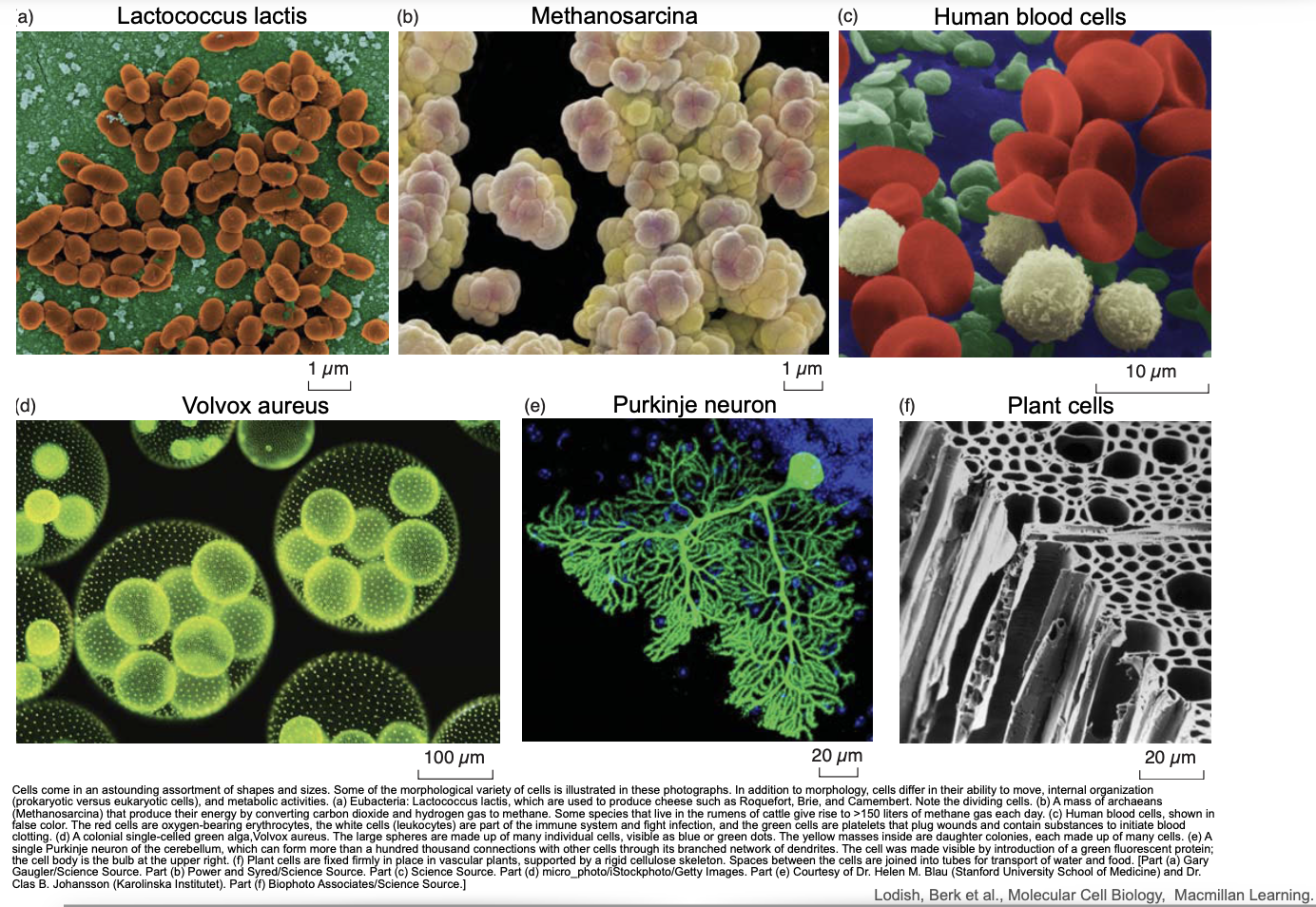
-
The document includes micrographs and descriptions of various cell types, highlighting the morphological and functional diversity of life:
-
Lactococcus lactis: A spherical bacterium used in cheese production; demonstrates simple prokaryotic division.
-
Methanosarcina: An archaeon that generates methane; illustrates metabolic specialization.
-
Human blood cells: Includes erythrocytes (oxygen transport), leukocytes (immune response), and platelets (clotting).
-
Volvox aureus: A multicellular green alga with reproductive colonies, demonstrating early forms of cellular cooperation.
-
Purkinje neuron: Highly branched cell in the cerebellum responsible for integrating large amounts of synaptic input.
-
Plant cells: Exhibit rigid cell walls, organized cytoplasm, and are interconnected in vascular tissues.
-
These examples reflect how structure relates to function and how evolutionary pressures have shaped cellular adaptations.
Universal Tree of Life
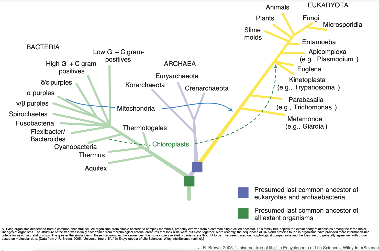
Phylogenetic Foundations
-
All life forms evolved from a common ancestor, likely a single-celled organism.
-
The tree is divided into three domains:
-
Bacteria: Prokaryotic cells with unique membrane and metabolic features.
-
Archaea: Prokaryotic, but genetically and biochemically distinct from bacteria; often extremophiles.
-
Eukaryota: Includes all multicellular organisms and unicellular eukaryotes; characterized by compartmentalization.
-
-
Molecular sequence data (e.g., rRNA genes, conserved proteins) is used to infer evolutionary relationships, replacing earlier morphology-based trees.
-
This concept emphasizes the shared biochemical core of life (e.g., use of DNA/RNA, ATP, lipid bilayers), even in vastly different organisms.
The Molecules of Life
Biological Macromolecules
-
The major classes include:
-
Proteins: Enzymes (e.g., glutamine synthetase), structural elements (e.g., cytoskeletal proteins), signaling molecules (e.g., hormones like insulin), and transporters (e.g., hemoglobin).
-
Nucleic Acids: DNA and RNA; store and transmit genetic information.
-
Lipids: Form membranes and participate in signaling.
-
Carbohydrates: Provide structural support and serve as energy sources.
-
Scale and Structure
-
Diagrams compare the size and shape of different molecules at the nanometer scale, e.g.:
-
Glutamine synthetase is shown next to a portion of a lipid bilayer and a DNA double helix.
-
Emphasizes how molecular dimensions affect interactions and localization in cellular space.
-
Principles of Molecular Interactions
Molecular Complementarity
-
Specific binding (e.g., enzyme-substrate, receptor-ligand) is determined by:
-
Structural fit (lock and key or induced fit models)
-
Chemical compatibility (e.g., hydrophobic, ionic, hydrogen bonds)
-
Polymerization
-
Monomers (e.g., nucleotides, amino acids) are covalently linked to form:
-
Polymers (e.g., DNA, proteins)
-
Directional polymers such as DNA (5′ → 3′) are critical for templated processes like replication and transcription.
-
Chemical Equilibrium
-
Reversible reactions are characterized by an equilibrium constant (Keq), which reflects the ratio of forward to reverse reaction rates (kf / kr).
-
Biological reactions are often maintained away from equilibrium to allow regulation and responsiveness.
Chemical Energy and ATP
-
The hydrolysis of ATP (adenosine triphosphate) provides free energy to drive endergonic reactions.
-
The energy comes from breaking phosphoanhydride bonds, releasing ADP and inorganic phosphate (Pi).
DNA Structure and Function
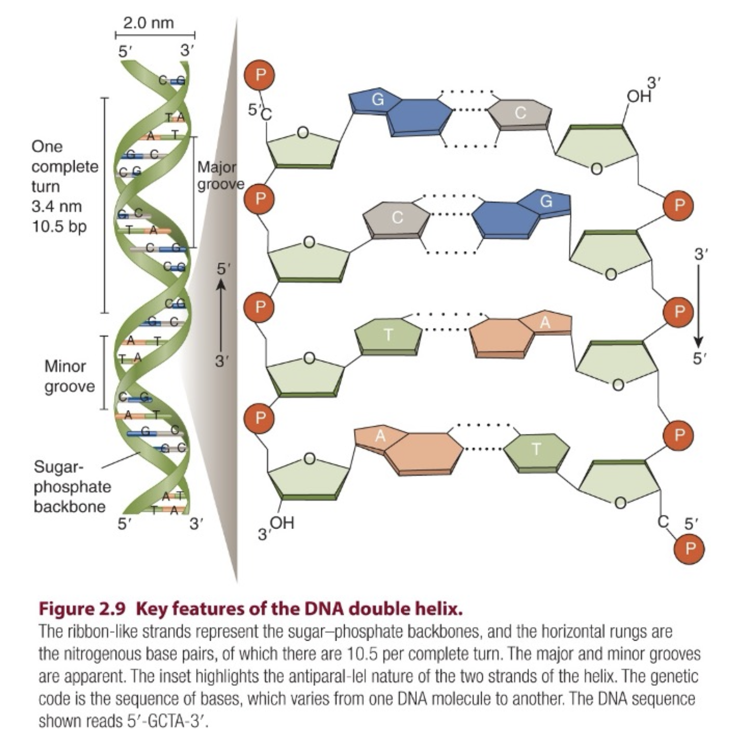
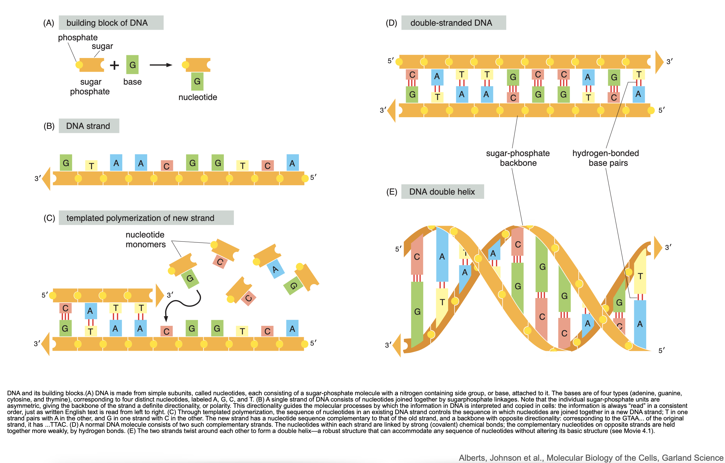
Building Blocks and Polarity
-
DNA is composed of four nucleotides: adenine (A), thymine (T), cytosine (C), guanine (G).
-
Each nucleotide contains:
-
A phosphate group
-
A deoxyribose sugar
-
A nitrogenous base
-
Double Helix and Base Pairing
-
Two complementary strands form a right-handed double helix.
-
Base pairing rules:
-
A pairs with T via 2 hydrogen bonds
-
G pairs with C via 3 hydrogen bonds
-
-
This structure allows for high-fidelity replication and robust information storage.
what is the difference between 5 prime and 3 prime on DNA helix?
The Central Dogma of Molecular Biology
Information Flow
-
Replication: Copying of DNA before cell division.
-
Transcription: Syxnthesis of RNA from DNA by RNA polymerase.
-
Translation: Synthesis of protein on ribosomes using mRNA as template.
Templated Polymerization
-
Each step involves using an existing strand (DNA or RNA) to guide the formation of a complementary strand or protein sequence.
-
Directionality is maintained (e.g., 5′ → 3′ in nucleic acid synthesis; N-terminal to C-terminal in proteins).
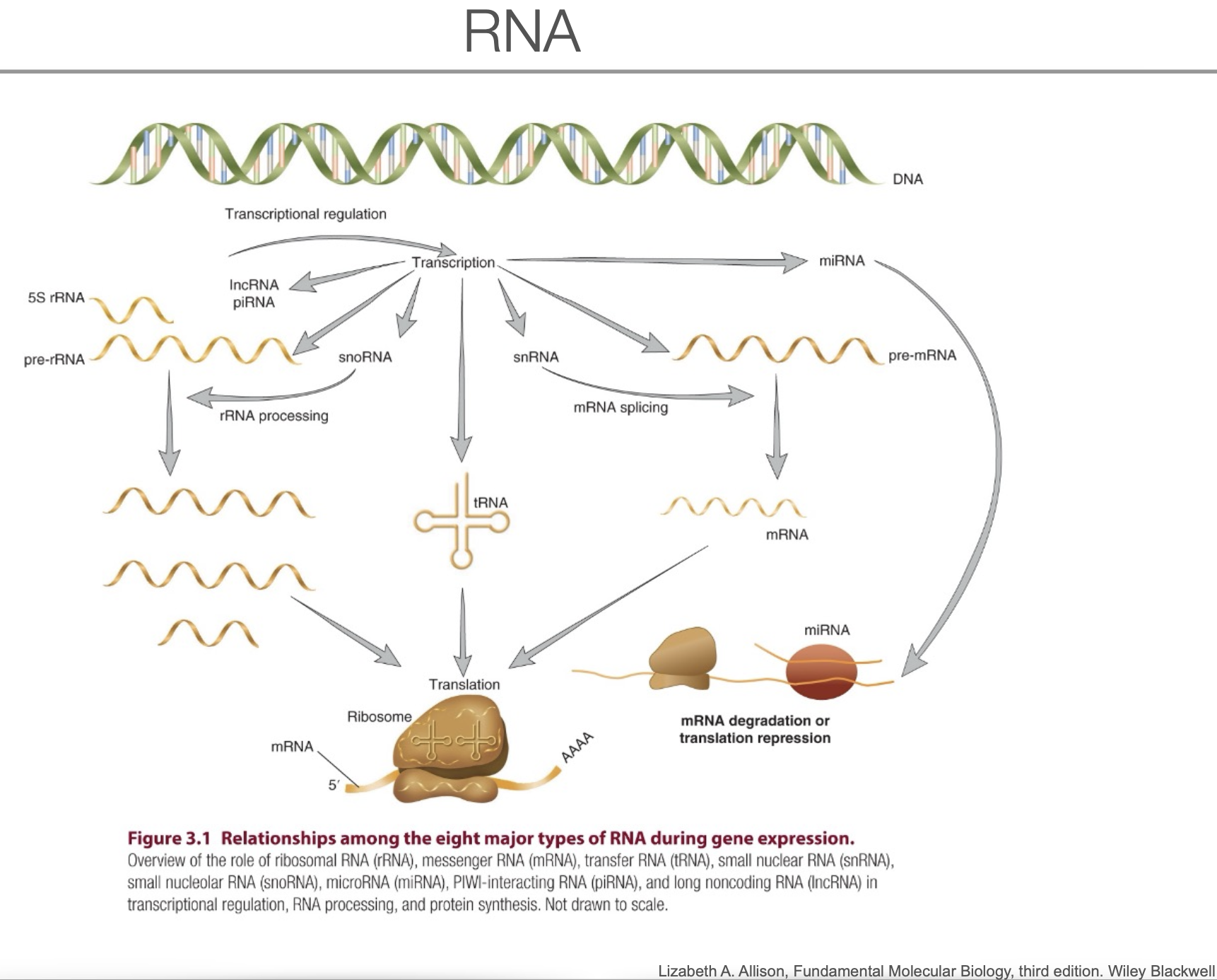
Transclude of Gene#gene-expression
Questions CNM 01_Introduction_1
Expanded Introduction (01_Introduction_2.pdf)**
What Is Molecular Biology?
-
Molecular biology aims to understand life in chemical and molecular terms.
-
It focuses on macromolecules—DNA, RNA, proteins—and their interactions.
-
The goal is to understand how these molecular systems give rise to cellular functions and organismal biology.
Scope of Molecular Biology
-
Originally focused on genetic information flow (DNA → RNA → protein).
-
Now expanded to include:
-
Gene regulation
-
Chromatin structure
-
RNA processing and modifications
-
Protein folding, trafficking, and degradation
-
Epigenetics and non-coding RNAs
-
The Central Dogma of Molecular Biology
DNA → RNA → Protein
-
DNA is transcribed into RNA.
-
RNA is processed (splicing, capping, polyadenylation).
-
mRNA is then translated into protein.
-
Some RNAs (e.g. rRNA, tRNA, miRNA) are not translated but serve functional roles.
Transcription Initiation in Eukaryotes
The Promoter Region
-
Contains key sequence motifs, including the TATA box, where RNA Polymerase II (Pol II) binds with the help of transcription factors.
-
TFIID, containing TATA-binding protein (TBP), binds first and initiates assembly of the pre-initiation complex (PIC).
General Transcription Factors (GTFs)
-
Core components of the transcription machinery:
-
TFIIA: stabilizes TBP-DNA interaction
-
TFIIB: bridges TBP and Pol II
-
TFIIF: escorts Pol II
-
TFIIE: recruits TFIIH
-
TFIIH: has helicase and kinase activity
-
Role of TFIIH
-
Unwinds DNA at the transcription start site (helicase activity).
-
Phosphorylates the C-terminal domain (CTD) of RNA Pol II.
- This releases Pol II from the promoter and initiates elongation.
The RNA Polymerase II Complex
Structure and Function
-
Multi-subunit enzyme that catalyzes 5′ → 3′ RNA synthesis.
-
Requires a DNA template and ribonucleotide triphosphates (rNTPs).
-
Cannot initiate transcription on its own—needs GTFs and chromatin access.
Alternative Splicing
Definition and Mechanism
-
A process that allows a single gene to produce multiple mRNA isoforms.
-
Performed by the spliceosome, a large RNA-protein complex composed of snRNPs:
-
U1, U2, U4, U5, and U6
-
Splice Site Recognition
-
Splicing depends on conserved sequences:
-
5′ splice site, branch point A, and 3′ splice site.
-
-
U1 binds the 5′ splice site, U2 binds the branch point, and the U4/U6.U5 tri-snRNP completes assembly.
-
Two transesterification reactions excise the intron and ligate exons.
Significance
-
Enables proteomic diversity from a limited number of genes.
-
Frequently regulated in a tissue- or activity-specific manner (e.g., neurons).
Protein Levels Are Not Just Transcription-Dependent
Translation and Degradation Matter
-
mRNA levels do not correlate linearly with protein levels.
-
Post-transcriptional regulation (e.g. RNA stability, translational efficiency) and protein degradation rates are crucial determinants of protein abundance.
RNA Secondary Structure
Functional Implications
-
RNA is single-stranded and can fold into complex 3D structures.
-
These structures affect:
-
Translation efficiency
-
Splicing
-
Localization
-
Interaction with RNA-binding proteins
-
Key Molecular Concepts Reinforced
-
Templated Polymerization
-
Used in DNA replication, transcription, and translation.
-
Ensures fidelity through base-pairing rules.
-
-
5′ to 3′ Directionality
-
All polymerases synthesize nucleic acids from 5′ to 3′.
-
Antiparallel nature of DNA necessitates complex machinery (e.g., Okazaki fragments in lagging strand synthesis).
-
-
CTD Phosphorylation
-
The C-terminal domain of RNA Pol II acts as a regulatory hub.
-
Different phosphorylation states recruit:
-
Capping enzymes
-
Splicing machinery
-
Polyadenylation factors
-
-
Summary Table – Splicing Overview
| Step | Molecular Component | Function |
|---|---|---|
| 1 | U1 snRNP | Binds 5′ splice site |
| 2 | U2 snRNP | Binds branch point A |
| 3 | U4/U6-U5 tri-snRNP | Assembles spliceosome |
| 4 | First transesterification | Cuts at 5′ splice site |
| 5 | Second transesterification | Ligates exons |
Conclusion
The PDF sets the foundation for understanding gene expression regulation with a focus on transcription initiation and RNA processing, both essential for neuronal identity, plasticity, and disease states. It underscores the importance of multi-layered regulation beyond transcription and introduces the molecular logic of modularity, directionality, and dynamic control in gene expression.
Questions CNM 01_Introduction_2
02_Introduction_3.pdf
🔬 The Chemical and Physical Principles Underlying Cell Biology
Cells Are Made of Molecules Governed by Physical and Chemical Laws
-
Biological systems obey the same laws of physics and chemistry that govern non-living systems.
-
Cellular organization and function are shaped by:
-
Thermodynamics
-
Kinetics
-
Electrostatics
-
Hydrophobic effects
-
-
These principles determine molecular assembly, stability, interaction, and reaction rates.
The Importance of Water
-
Cells exist in an aqueous environment, and water is:
-
A solvent for ions and polar molecules.
-
A participant in hydrolysis and condensation reactions.
-
-
Hydrophobic and hydrophilic interactions are key forces driving membrane formation, protein folding, and molecular recognition.
⚛️ Chemical Bonds and Molecular Forces
Covalent Bonds
-
Formed by the sharing of electrons between atoms.
-
Examples:
-
C–C, C–H, C–N, C–O.
-
-
Provide molecular backbone stability.
Non-Covalent Interactions
Include:
-
Ionic bonds (electrostatic attraction between oppositely charged ions)
-
Hydrogen bonds (important for base pairing and protein structure)
-
Van der Waals interactions (transient dipole-induced forces)
-
Hydrophobic interactions (entropy-driven organization of nonpolar substances)
These interactions:
-
Are weaker individually than covalent bonds.
-
Drive dynamic molecular recognition, such as enzyme-substrate or receptor-ligand binding.
🧬 Biomolecules in Cells
Proteins
-
Polymers of amino acids, joined by peptide bonds.
-
Fold into specific 3D structures dictated by amino acid sequence.
-
Functions include:
-
Enzymes
-
Receptors
-
Structural components
-
Transporters
-
Signaling molecules
-
Nucleic Acids
-
DNA and RNA are polymers of nucleotides, linked by phosphodiester bonds.
-
Nucleotide = base (A, T/U, G, C) + ribose/deoxyribose + phosphate.
-
DNA is double-stranded and stores hereditary information.
-
RNA is single-stranded and plays roles in:
-
Protein synthesis (mRNA, rRNA, tRNA)
-
Gene regulation (miRNA, siRNA)
-
Carbohydrates
-
Sugars and their polymers (e.g. glycogen, cellulose).
-
Functions:
-
Energy storage (glucose, glycogen)
-
Structural support (cell walls)
-
Cell-cell recognition (glycoproteins, glycolipids)
-
Lipids
-
Diverse group: fatty acids, phospholipids, cholesterol, triglycerides.
-
Functions:
-
Form biological membranes
-
Store energy
-
Act as signaling molecules (e.g. steroid hormones)
-
🧪 Molecular Complementarity and Binding
Molecular Complementarity
-
Specific fit between molecules (e.g. enzyme and substrate, antibody and antigen) depends on:
-
Shape
-
Charge
-
Hydrogen bonding
-
-
It underlies all specific molecular recognition in biology.
Binding Affinity and Specificity
-
Strength and specificity of molecular interactions depend on:
-
The number and types of non-covalent bonds.
-
The geometry of interaction surfaces.
-
-
High affinity ≠ high specificity (e.g. some molecules bind tightly but to many targets).
🌡️ Thermodynamics and Kinetics
Free Energy and Reaction Spontaneity
-
A reaction is spontaneous if it decreases Gibbs free energy (ΔG < 0).
-
ATP hydrolysis is a central energy-releasing reaction in cells.
Activation Energy and Catalysis
-
Reactions require overcoming an energy barrier (Ea).
-
Enzymes:
-
Lower the activation energy.
-
Do not change ΔG.
-
Greatly accelerate reaction rates.
-
🧬 Templated Polymerization
-
All major biological macromolecules (DNA, RNA, proteins) are made by templated polymerization:
-
DNA → RNA (transcription)
-
RNA → Protein (translation)
-
-
Template-directed base-pairing ensures fidelity of information transfer.
📌 Summary
| Biomolecule | Building Block | Bond Type | Major Functions |
|---|---|---|---|
| Protein | Aminosäure, amino acid | Peptide | Catalysis, structure, signaling |
| DNA/RNA | nucleotides | Phosphodiester | Genetic information, translation |
| Carbohydrates | Sugars | Glycosidic | Energy, structure, recognition |
| Lipids | Fatty acids, etc. | Hydrophobic interactions | Membranes, energy storage, signaling |
Questions CNM 02_Introduction_3
Cellular and Molecular Neuroscience – Cellular Components II (
04_Cellular_components.pdf
)
🧠 Recap: Glial Cell Functions in the Nervous System
Four Main Functions of Glial Cells
-
Homeostasis
-
Glia regulate extracellular ion concentrations, especially K⁺.
-
Remove excess neurotransmitters (e.g., glutamate) from synaptic clefts.
-
Control pH and metabolite levels.
-
-
Myelination
-
Oligodendrocytes (CNS) and Schwann cells (PNS) insulate axons.
-
Myelin increases conduction velocity and reduces energy costs.
-
Defects in myelination lead to neurological disorders (e.g., MS).
-
-
Immune Defense
-
Microglia act as the brain’s immune cells.
-
They remove apoptotic cells, debris, and infectious agents.
-
Involved in neuroinflammation and disease progression.
-
-
Modulation of Neural Activity
-
Astrocytes influence synaptic transmission via neurotransmitter uptake and gliotransmission.
-
They can release gliotransmitters (e.g., D-serine, ATP) and modulate neuronal excitability.
-
🧬 Wiring the Brain: Developmental Mechanisms
Myelination Timing and Regional Differences
-
Myelination occurs postnatally and follows a spatiotemporal gradient:
-
Begins in sensory and motor pathways.
-
Later in association cortices (prefrontal cortex, involved in cognition).
-
-
Continues into early adulthood (e.g., age 25+ in humans).
-
Experience-dependent: learning and stimulation influence myelin growth.
Evidence for Experience-Dependent Myelination
-
Examples:
-
Animals raised in enriched environments show increased myelination.
-
Learning new motor tasks (e.g., running wheels) can enhance myelin thickness.
-
-
Activity can influence oligodendrocyte precursor differentiation.
🧪 Plasticity Beyond Neurons
Glial Plasticity
-
Astrocytes and oligodendrocytes are not static support cells:
-
Astrocytes undergo morphological changes in response to neuronal activity.
-
Oligodendrocyte function is modulated by electrical activity and learning.
-
Functional Implications
-
Glial plasticity is linked to:
-
Learning and memory
-
Adaptive changes in neural circuits
-
Repair mechanisms after injury
-
🧩 Clinical Implications of Glial Dysfunction
Neurological and Psychiatric Disorders
-
Glial abnormalities are implicated in:
-
Multiple Sclerosis (demyelination)
-
Alzheimer’s Disease (inflammation, astrocytic dysfunction)
-
Depression and Schizophrenia (astrocyte and oligodendrocyte involvement)
-
Targeting Glia for Therapy
-
Emerging approaches:
-
Stimulating remyelination via oligodendrocyte progenitors
-
Modulating astrocytic glutamate uptake to prevent excitotoxicity
-
Controlling microglial activation to reduce chronic inflammation
-
Summary Table – Functions and Features of Glial Cells
| Glial Cell Type | Key Functions | Notes |
|---|---|---|
| Astrocytes | Homeostasis, synaptic modulation, blood-brain barrier | Respond to neuronal activity |
| Oligodendrocytes | CNS myelination | One cell → multiple axons |
| Schwann Cells | PNS myelination | One cell → one axon segment |
| Microglia | Immune surveillance, synaptic pruning | Dynamic responders to damage/disease |
**### ⚡ Cellular and Molecular Neuroscience – Ionotropic Receptors
🧠 Overview: What Are Ionotropic Receptors?
Ionotropic receptors are ligand-gated ion channels that mediate fast synaptic transmission in the nervous system. Upon neurotransmitter binding, they undergo conformational changes that allow ions to flow across the membrane, leading to depolarization or hyperpolarization of the postsynaptic neuron.
⇒ Ionotropic Receptors
🔑 Key Properties
Speed and Mechanism
-
Fast activation: Onset within milliseconds.
-
Direct ion flow: Neurotransmitter binding opens the pore, allowing Na⁺, K⁺, Ca²⁺, or Cl⁻ to pass, depending on the receptor type.
-
Reversible and transient: Channels open only while ligand is bound.
Contrast with Metabotropic Receptors
-
Ionotropic: direct, fast, local
-
Metabotropic: indirect, slower, modulatory (via G proteins)
🧬 Structural Organization
Subunit Composition
-
Typically tetrameric or pentameric assemblies of homologous subunits.
-
Each subunit contributes to the ion pore and ligand-binding domain.
-
Extracellular domain: binds neurotransmitter.
-
Transmembrane domain: forms ion-conducting pore.
Structural Families
-
Cys-loop receptors (e.g., GABA_A, glycine, nicotinic AChR)
-
Ionotropic glutamate receptors (AMPA, NMDA, kainate)
-
ATP-gated channels (P2X receptors)
🔥 Major Classes of Ionotropic Receptors
1.
Glutamate Receptors
AMPA Receptors
-
Mediate fast excitatory transmission.
-
Permeable to Na⁺ and K⁺, sometimes Ca²⁺.
-
Rapid kinetics; important in baseline synaptic activity.
NMDA Receptors
-
Also glutamate-gated, but with unique properties:
-
Voltage-dependent block by Mg²⁺.
-
Requires co-agonist (glycine or D-serine).
-
Permeable to Ca²⁺, linking to plasticity mechanisms.
-
-
Central in synaptic plasticity, learning, and memory.
Kainate Receptors
-
Less understood.
-
Modulate both excitatory and inhibitory transmission.
2.
GABA_A Receptors
-
Main inhibitory receptor in the CNS.
-
Permeable to Cl⁻ ions.
-
Activation typically causes hyperpolarization.
-
Composed of α, β, γ, and other subunits.
-
Target for:
-
Benzodiazepines
-
Barbiturates
-
Alcohol
-
General anesthetics
-
3.
Glycine Receptors
-
Also Cl⁻-permeable inhibitory receptors.
-
Prominent in spinal cord and brainstem.
-
Share structural similarity with GABA_A receptors.
4.
Nicotinic Acetylcholine Receptors (nAChR)
-
Excitatory receptors permeable to Na⁺ and K⁺.
-
Found in:
-
Neuromuscular junctions
-
Autonomic ganglia
-
CNS circuits
-
-
Targeted by:
-
Nicotine
-
Curare
-
α-bungarotoxin
-
5.
ATP-Gated P2X Receptors
-
Activated by extracellular ATP.
-
Non-selective cation channels.
-
Involved in:
-
Pain signaling
-
Inflammation
-
Neuromodulation
-
🧪 Pharmacological Relevance
Modulation by Drugs
-
Ionotropic receptors are key drug targets:
-
Anesthetics enhance GABA_A function.
-
Antiepileptics target excitatory/inhibitory balance.
-
Ketamine blocks NMDA receptors (used as anesthetic and antidepressant).
-
Toxins and Antagonists
-
Snake toxins (e.g., α-bungarotoxin) block nAChRs.
-
CNQX, APV, and MK-801 are glutamate receptor antagonists used in research.
🧩 Functional Implications
Synaptic Plasticity
-
NMDA and AMPA receptor dynamics underlie LTP (long-term potentiation) and LTD (long-term depression).
-
Receptor trafficking, phosphorylation, and subunit composition regulate plasticity.
Development and Disease
-
Ionotropic receptor dysfunction is implicated in:
-
Epilepsy
-
Schizophrenia
-
Neurodegeneration
-
Chronic pain
-
Summary Table – Major Ionotropic Receptors
| Receptor | Ions Conducted | Function | Antagonists/Modulators |
|---|---|---|---|
| AMPA | Na⁺, K⁺ (±Ca²⁺) | Fast excitation | CNQX |
| NMDA | Na⁺, K⁺, Ca²⁺ | Plasticity | APV, MK-801, ketamine |
| GABA_A | Cl⁻ | Inhibition | Bicuculline, benzodiazepines |
| Glycine | Cl⁻ | Inhibition | Strychnine |
| nAChR | Na⁺, K⁺ | Excitation | Curare, α-bungarotoxin |
| P2X | Na⁺, K⁺, Ca²⁺ | Modulation | PPADS |
06_Neurotransmission.pdf
🔄 Synaptic Transmission: Overview
Neurotransmission is the core mechanism by which neurons communicate, using chemical synapses to transmit information across synaptic clefts. The process involves:
-
Action potential arrival at the axon terminal.
-
Calcium influx via voltage-gated Ca²⁺ channels.
-
Vesicle fusion and neurotransmitter release.
-
Binding of neurotransmitters to postsynaptic receptors.
-
Signal termination via reuptake, enzymatic degradation, or diffusion.
🧠 Morphological and Functional Organization
Types of Synapses
-
Electrical Synapses:
-
Direct ionic current via gap junctions.
-
Bidirectional and fast (no synaptic delay).
-
-
Chemical Synapses:
-
Involve neurotransmitters crossing synaptic clefts.
-
Unidirectional with synaptic delay (~1 ms).
-
Can be excitatory or inhibitory depending on receptor type.
-
Active Zones
-
Specialized presynaptic regions where:
-
Docked vesicles are primed for release.
-
Voltage-gated Ca²⁺ channels are localized.
-
SNARE machinery is preassembled.
-
🧬 Molecular Machinery of Vesicle Fusion
1.
Docking
-
Vesicles are transported to the active zone and tethered near the membrane.
-
Involves Rab3 and RIM proteins to position vesicles close to Ca²⁺ channels.
2.
Priming
-
SNARE complex partially assembles:
-
Synaptobrevin/VAMP (vesicle membrane)
-
Syntaxin + SNAP-25 (plasma membrane)
-
-
Requires Munc13 and Munc18 for proper SNARE configuration.
3.
Fusion and Exocytosis
-
Triggered by Ca²⁺ influx, sensed by synaptotagmin.
-
Synaptotagmin binds Ca²⁺ and displaces complexin, enabling full SNARE “zippering.”
-
This drives membrane fusion, forming a fusion pore for neurotransmitter release.
🔋 Calcium Dependence of Release
-
Release probability scales steeply with intracellular Ca²⁺ concentration (cooperative binding).
-
Spatial coupling of Ca²⁺ channels and vesicles is critical (nanodomain coupling).
-
Different vesicles may respond to different Ca²⁺ dynamics.
🧠 Functional Modes of Release
Synchronous Release
-
Tight temporal coupling with the action potential.
-
Requires high local Ca²⁺ and is mediated primarily by synaptotagmin-1.
Asynchronous Release
-
Delayed, often due to residual Ca²⁺ or lower-affinity sensors (e.g., synaptotagmin-7).
-
Modulates firing patterns or synaptic strength.
Spontaneous Release
-
Occurs without action potentials.
-
Provides background synaptic noise and homeostatic tone.
🧪 Endocytosis and Vesicle Recycling
After exocytosis:
-
Vesicle membrane and proteins are retrieved via:
-
Clathrin-mediated endocytosis (dominant mode).
-
Kiss-and-run (hypothesized rapid mechanism).
-
-
Key proteins: dynamin, clathrin, AP-2.
-
Vesicles are refilled with neurotransmitter via vacuolar-type ATPases and transporters.
🧩 Regulation and Plasticity
-
Release can be modulated by:
-
Presynaptic receptors (e.g., autoreceptors, heteroreceptors).
-
Short-term plasticity (facilitation, depression).
-
Long-term changes (e.g., changes in release probability or vesicle pool size).
-
🧬 Experimental Tools for Studying Neurotransmission
Electrophysiological Approaches
-
Patch clamp: measures synaptic currents.
-
Capacitance measurements: detect vesicle fusion via membrane surface changes.
Optical Methods
-
FM dyes: label recycling vesicles.
-
pHluorins: genetically encoded indicators of vesicle fusion.
-
Calcium imaging: tracks Ca²⁺ transients and sensor recruitment.
Genetic Manipulations
-
Knockouts of SNARE proteins, synaptotagmin, complexin to dissect release machinery.
-
Use of toxins (e.g., tetanus, botulinum) to block specific SNAREs.
🧠 Clinical and Pharmacological Relevance
-
Botulinum and tetanus toxins cleave SNARE proteins, blocking neurotransmission (paralysis or spasticity).
-
Disorders involving altered neurotransmitter release:
-
Schizophrenia
-
Epilepsy
-
Parkinson’s disease
-
-
Modulation of release is a target for:
-
Antiepileptics
-
Anxiolytics
-
Anesthetics
-
Summary Table – Core Steps of Neurotransmitter Release
| Step | Key Proteins | Function |
|---|---|---|
| Docking | Rab3, RIM | Vesicle tethering at active zone |
| Priming | Munc13, Munc18, SNAREs | Prepare vesicle for fusion |
| Fusion | SNAREs, synaptotagmin | Ca²⁺-dependent membrane fusion |
| Endocytosis | Clathrin, dynamin | Vesicle recycling and protein retrieval |
Here is a deep and detailed academic summary of your uploaded file 07_Ionotropic receptors.pdf, organized with , , and headings to reflect the structure and depth of the material.
07_Ionotropic receptors.pdf
🧠 What Are Ionotropic Receptors?
Ionotropic receptors are ligand-gated ion channels that mediate fast synaptic transmission. Upon neurotransmitter binding, they undergo conformational changes that open an ion-conducting pore, allowing rapid flux of specific ions and immediate changes in membrane potential.
🧬 Structural Features of Ionotropic Receptors
General Architecture
-
Multimeric complexes, usually formed by 4–5 subunits.
-
Each subunit has:
-
Extracellular domain (ECD): binds neurotransmitter.
-
Transmembrane domain (TMD): forms ion-conducting pore (usually 4 membrane-spanning regions per subunit).
-
Some subtypes have intracellular loops involved in regulatory signaling or anchoring to scaffolding proteins.
-
Ion Selectivity and Gating
-
Determined by:
-
Pore size and shape
-
Charge distribution at selectivity filter
-
-
Can be cation-selective (e.g. Na⁺, K⁺, Ca²⁺) or anion-selective (e.g. Cl⁻).
🔑 Major Classes of Ionotropic Receptors
1.
Cys-loop Receptors
-
Named after conserved disulfide bridge (cys-loop) in extracellular domain.
-
Pentameric structure (5 subunits).
-
Includes:
-
GABA_A receptors
-
Glycine receptors
-
Nicotinic acetylcholine receptors (nAChRs)
-
5-HT₃ receptors
-
GABA
A
Receptors
-
Main inhibitory receptors in CNS.
-
Permeable to Cl⁻ ions.
-
Composed of various combinations of α, β, γ, δ subunits.
-
Target of benzodiazepines, barbiturates, anesthetics, and ethanol.
Glycine Receptors
-
Also Cl⁻-permeable.
-
Mediate inhibition in spinal cord and brainstem.
-
Target of strychnine (a convulsant toxin).
nAChRs
-
Permeable to Na⁺ and K⁺, sometimes Ca²⁺.
-
Mediate fast excitation at neuromuscular junctions and in CNS.
-
Blocked by curare, α-bungarotoxin.
2.
Ionotropic Glutamate Receptors
-
Tetrameric structure.
-
Three major types:
-
AMPA receptors
-
NMDA receptors
-
Kainate receptors
-
AMPA Receptors
-
Fastest excitatory receptors.
-
Na⁺ and K⁺ permeable.
-
GluA2 subunit editing regulates Ca²⁺ permeability.
-
Rapid kinetics and essential for baseline synaptic transmission.
NMDA Receptors
-
Require both glutamate and co-agonist (glycine or D-serine).
-
Blocked by Mg²⁺ at resting potential—need depolarization to open.
-
Permeable to Na⁺, K⁺, and Ca²⁺.
-
Act as coincidence detectors for synaptic plasticity (LTP/LTD).
-
Targeted by APV, MK-801, ketamine.
Kainate Receptors
-
Similar to AMPA but with slower kinetics and more nuanced roles.
-
Found in pre- and postsynaptic compartments.
-
Modulate network excitability.
3.
P2X Receptors
-
ATP-gated ion channels.
-
Non-selective for cations (Na⁺, K⁺, Ca²⁺).
-
Play roles in:
-
Pain
-
Inflammation
-
Purinergic signaling
-
-
Trimeric structure.
-
Blocked by antagonists like PPADS.
📈 Functional Principles of Ionotropic Receptors
Speed
-
Activated within milliseconds of ligand binding.
-
Direct ion flow leads to immediate postsynaptic potentials.
Direction of Effect
-
Excitatory if net effect is depolarization (e.g., AMPA, nAChR).
-
Inhibitory if net effect is hyperpolarization or shunting inhibition (e.g., GABA_A, glycine).
Desensitization
-
Receptors can enter an inactive state despite ligand presence.
-
Important for controlling signal duration and preventing overexcitation.
🧠 Integration in Neural Circuits
-
Ionotropic receptors are synapse-localized, enabling:
-
Spatially confined, rapid signaling.
-
Specificity based on subunit composition and receptor localization.
-
-
Their properties (e.g., decay time, ion permeability) are critical in:
-
Temporal coding
-
Synaptic plasticity
-
Excitation/inhibition balance
-
🧪 Pharmacological Modulation
| Receptor | Agonists | Antagonists / Modulators |
|---|---|---|
| AMPA | Glutamate, AMPA | CNQX, NBQX |
| NMDA | Glutamate + Gly/D-Serine | APV, MK-801, ketamine |
| GABA_A | GABA | Bicuculline, picrotoxin |
| Glycine | Glycine | Strychnine |
| nAChR | Acetylcholine, nicotine | α-bungarotoxin, curare |
| P2X | ATP | PPADS, suramin |
🧬 Summary
| Receptor Family | Subtype | Ion Flow | Synaptic Role |
|---|---|---|---|
| Cys-loop | GABA_A | Cl⁻ | Fast inhibition |
| Glycine | Cl⁻ | Fast inhibition in spinal cord | |
| nAChR | Na⁺, K⁺ | Fast excitation | |
| Glutamate | AMPA | Na⁺, K⁺ (±Ca²⁺) | Fast excitation |
| NMDA | Na⁺, K⁺, Ca²⁺ | Plasticity, coincidence detection | |
| Kainate | Na⁺, K⁺ | Excitation, modulation | |
| P2X | P2X1–7 | Na⁺, K⁺, Ca²⁺ | Pain, inflammation, modulation |
08_Metabotropic receptors.pdf
📚 What Are Metabotropic Receptors?
Metabotropic receptors are G protein-coupled receptors (GPCRs) that, unlike ionotropic receptors, do not form ion channels. Instead, they trigger intracellular signaling cascades via G-proteins, resulting in modulatory, slower, and longer-lasting effects on neuronal function. They are essential for plasticity, learning, neuromodulation, and homeostatic regulation.
6
🧬 Structural and Molecular Characteristics
General Structure
-
Single polypeptide spanning the membrane seven times (7TM).
-
An extracellular domain binds neurotransmitter (ligand).
-
An intracellular domain interacts with G proteins.
G-Protein Coupling
-
Receptors activate heterotrimeric G-proteins made of:
-
α-subunit (defines class: Gs, Gi/o, Gq/11)
-
βγ dimer
-
-
Binding of ligand to receptor → conformational change → exchange of GDP for GTP on α → α and βγ subunits separate and activate downstream effectors.
🔄 Signal Transduction Pathways
G
s
Pathway
-
Stimulates adenylate cyclase → ↑ cAMP → activates PKA.
-
Example receptors: D1 dopamine, β-adrenergic, some serotonin receptors.
G
i/o
Pathway
-
Inhibits adenylate cyclase → ↓ cAMP.
-
βγ subunit can:
-
Open GIRK (K⁺) channels → hyperpolarization.
-
Inhibit voltage-gated Ca²⁺ channels → ↓ transmitter release.
-
-
Example: GABAB, D2 dopamine, α2-adrenergic.
G
q/11
Pathway
-
Activates phospholipase C (PLC) → cleaves PIP₂ into:
-
DAG → activates PKC
-
IP₃ → triggers Ca²⁺ release from ER
-
-
Example: mGluR1/5, M1/M3 muscarinic, α1-adrenergic
🧪 Key Metabotropic Receptor Classes in Neuroscience
1.
Metabotropic Glutamate Receptors (mGluRs)
-
Divided into three groups:
-
Group I (mGluR1,5): Gq-coupled, increase Ca²⁺ and excitability.
-
Group II (mGluR2,3): Gi/o-coupled, reduce transmitter release.
-
Group III (mGluR4,6,7,8): also Gi/o, often presynaptic autoreceptors.
-
-
Functions:
-
Synaptic plasticity (LTD)
-
Presynaptic modulation
-
Neuroprotection
-
2.
GABAB Receptors
-
Obligatory heterodimers: GABAB1 (binds ligand) + GABAB2 (couples to G-protein).
-
Gi/o-coupled:
-
Inhibits adenylate cyclase.
-
Opens K⁺ channels (postsynaptic inhibition).
-
Closes Ca²⁺ channels (presynaptic inhibition).
-
-
Found at pre- and postsynaptic sites.
-
Role: slow inhibition, shaping network oscillations and preventing overexcitation.
3.
Muscarinic Acetylcholine Receptors (mAChRs)
-
Subtypes:
-
M1, M3, M5: Gq, increase Ca²⁺ → excitatory modulation.
-
M2, M4: Gi/o, reduce cAMP and hyperpolarize neurons.
-
-
Roles:
-
Cortical excitability, attention, REM sleep, parasympathetic signaling
-
4.
Monoamine GPCRs
-
Dopamine Receptors:
-
D1-like (D1, D5): Gs, increase cAMP → facilitate LTP.
-
D2-like (D2, D3, D4): Gi/o, suppress cAMP → decrease excitability.
-
Roles in: reward, motivation, movement, learning
-
-
Serotonin (5-HT) Receptors:
-
5-HT1, 2, 4, 5, 6, 7: mostly GPCRs.
-
Roles in: mood, arousal, appetite, sensory processing
-
-
Adrenergic Receptors:
-
α1: Gq (vasoconstriction, arousal)
-
α2: Gi (inhibitory feedback)
-
β1–3: Gs (alertness, cardiac output, plasticity)
-
🧠 Functional Roles of Metabotropic Receptors
| Function | Mechanism |
|---|---|
| Neuromodulation | Changes the excitability of neurons based on brain state (e.g. attention) |
| Plasticity | Involved in LTD, gene expression, spine remodeling |
| Presynaptic regulation | Controls neurotransmitter release via Ca²⁺ channel inhibition |
| Network tuning | Shapes network rhythms and filtering (e.g. theta/gamma oscillations) |
🔁 Desensitization and Termination
-
GPCRs undergo desensitization via:
-
Phosphorylation by GRKs (G-protein receptor kinases)
-
Binding of β-arrestins → internalization and recycling/degradation
-
-
RGS proteins accelerate GTP hydrolysis → inactivate Gα subunit
✅ Summary: Ionotropic vs. Metabotropic Receptors
| Feature | Ionotropic | Metabotropic |
|---|---|---|
| Speed | Fast (ms) | Slow (100 ms – minutes) |
| Mechanism | Direct ion flow | G-protein signaling |
| Receptor structure | Multimeric channels | 7-transmembrane GPCR |
| Effect duration | Brief | Sustained |
| Functional domain | Point-to-point synaptic | Local and volume transmission |
| Synaptic role | Fast EPSP/IPSP | Modulation, plasticity, tone |
Let me know if you’d like:
-
A receptor family map (mGluRs, GABA_B, monoamines)
-
A flowchart from ligand binding to gene expression
-
30 study questions based on this summary
see also
Tags: neuroscience science
Superlink: 050 🧠Neuroscience
PCR
NGS
Questions for CMN
Source
Created: 03-04-25 14:07