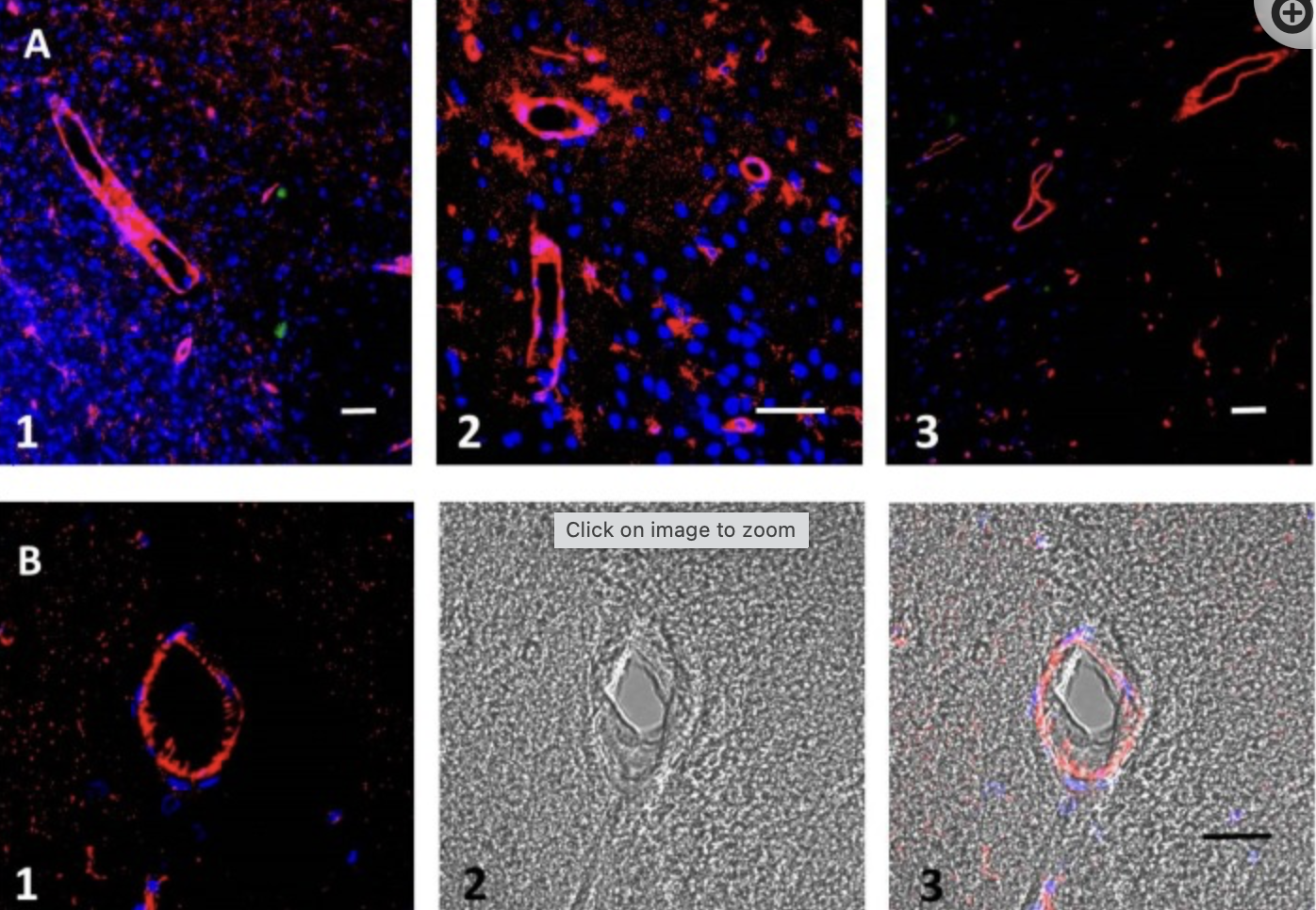
(A) Brain zone affected by ischemia. Amyloid beta (Aβ) peptide oligomer immunostaining (red), Congo red staining specific for aggregated amyloid (green). The walls of both large and small vessels are visible. Nuclear DAPI staining (blue) shows that Aβ does not mainly coincide with cells, whereas red staining is mainly present in blood vessels. Congo red staining specific for aggregated amyloid (green) is present as low-density spots in the tissue and not associated with any nucleus. (B) Cross-section of a blood vessel in the affected zone after stroke: (1) fluorescence, (2) bright field, (3) merged view. Scale bars, 100 µm**.**
It is known that beta-amyloid(Aβ) can form nonselective ion channels in the external cellular membrane of brain cells, producing dyshomeostasis, synaptic dysfunction, and osmotic changes leading to edema and cell death. If Aβ generated during ischemic stroke can damage neurons and other brain cells, blocking these channels might help cells to survive and to reduce swelling.
see also
Tags: neurobiology science
Superlink: 051 ☣Neurobiology 050 🧠Neuroscience
Memory in Sleep
Source
Martins, A. H., Zayas-Santiago, A., Ferrer-Acosta, Y., Martinez-Jimenez, S. M., Zueva, L., Diaz-Garcia, A., & Inyushin, M. (2019). Accumulation of Amyloid Beta (Aβ) Peptide on Blood Vessel Walls in the Damaged Brain after Transient Middle Cerebral Artery Occlusion. Biomolecules, 9(8). https://doi.org/10.3390/biom9080350
Created: 02-02-24 14:36