Phototransduction
chatbot
Phototransduction is a complex process that converts light into electrical signals in the eye, enabling vision. Here’s a simplified breakdown with key terms highlighted:
The Process of Phototransduction
-
Phototransduction: The biochemical process by which photoreceptors (rods and cones) in the retina convert light into electrical signals.
-
Rods and Cones: Specialized photoreceptor cells in the retina.
- Rods are highly sensitive to light and essential for night vision but do not distinguish colors.
- Cones are responsible for color vision and high visual acuity, functioning best in bright light.
Step 1:
- 11-cis retinal: A photosensitive molecule bound to opsin in photoreceptors. Light exposure converts it to all-trans retinal, initiating the phototransduction cascade.
Step 2:
-
Opsin therefore undergoes a conformational change to metarhodopsin II.
-
Rhodopsin: A protein in Rods composed of opsin and 11-cis retinal. Acts as a receptor that changes conformation when it absorbs light, starting the signal transduction process.
Step 3:
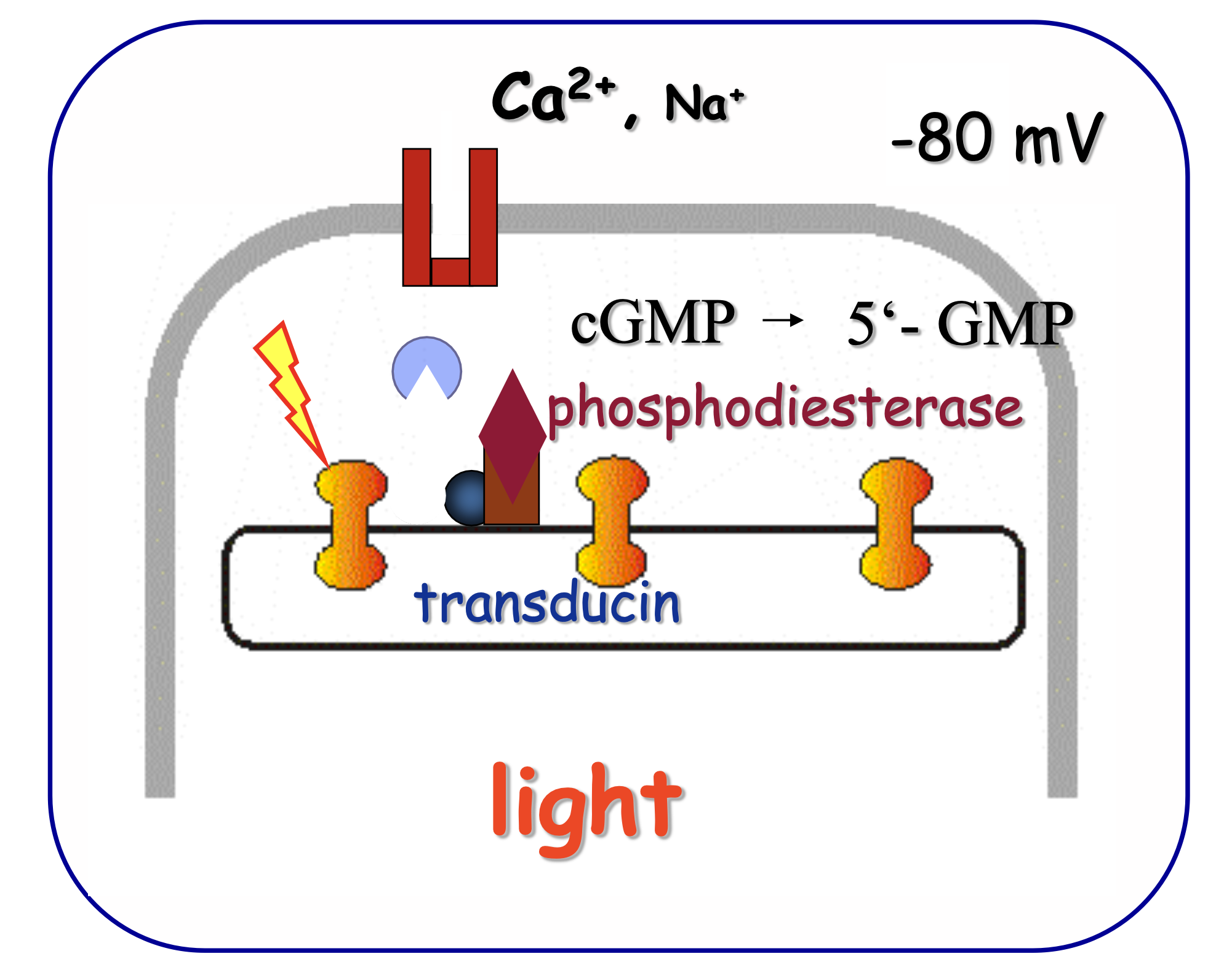
- Transducin: A G-protein activated by the conformational change in rhodopsin. It plays a crucial role in the signal cascade by activating the next molecule in the pathway.
Step 4:
-
Phosphodiesterase (PDE): An enzyme activated by transducin. It breaks down cGMP into 5‘-GMP, a crucial step in the phototransduction cascade.
-
cGMP (cyclic guanosine monophosphate): A second messenger molecule that, in the dark, binds to and keeps CNG-gated ion channels open, allowing cations to flow into the cell and depolarize it.
Step 5:
- CNG-gated ion channels: Channels that allow cations (calcium and sodium) to enter the photoreceptor cells. Closing of these channels in response to light leads to hyperpolarization of the cell membrane.
Signal Amplification
![]()
Terms
-
Hyperpolarization: A change in a cell’s membrane potential that makes it more negative. In photoreceptors, this stops the release of the neurotransmitter glutamate, altering the signal sent to bipolar cells.
-
Glutamate: A neurotransmitter released by photoreceptors in the dark. It has different effects on bipolar cells, depending on their type (on or off). Glutamate
-
Bipolar cells: Neurons in the retina that convey signals from the photoreceptors to the retinal ganglion cells. They are differentiated by their response to glutamate:
- On-bipolar cells have metabotropic glutamate receptors and become depolarized (activated) in the light when glutamate release is reduced.
- Off-bipolar cells have ionotropic glutamate receptors and become hyperpolarized (inhibited) in the light.
⇒ bipolar cells
-
Retinal Ganglion Cells (RGCs): Neurons that receive signals from bipolar cells and transmit them to the brain via the optic nerve. Their activity is modulated by the state of bipolar cells, leading to the perception of light.
⇒ retinal ganglion cells
This process allows the visual system to convert light into a neural signal that the brain can interpret, enabling sight.
![]()
Slides
- light induces conversion of 11-cis into all-trans retinal
- conformational change of rhodopsin causes dissociation and release of transducin
- transducin activates phosphodiesterase
- phosphodiesterase splits cGMP into 5‘-GMP
- CNG-gated ion channels close
- membrane of outer segment gets hyperpolarized
⇒ Sodium and CNG channels
In the dark
- photoreceptors are depolarized in the darkness
- they release the Neurotransmitter Glutamate
- on-bipolar cells possess inhibitory glutamate receptors, i.e. they become hyperpolarized
- connected retinal ganglion cells reduce their firing frequency („off- response“)
what also happens:
3. • off-bipolar cells possess excitatory glutamate receptors, i.e. they become depolarized by glutamate
4. connected retinal ganglion cells increase their firing frequency („on- response“)
back in the light
- photoreceptors are hyperpolarized in the light
- they no longer release the neurotransmitter glutamate
- on-bipolar cells return to a depolarized state
- connected retinal ganglion cells increase their firing frequency („on- response“)
what also happens:
3. off-bipolar cells return to a hyperpolarized state
4. connected retinal ganglion cells decrease their firing frequency („off- response“)
Phototransduction, the process by which light is converted into electrical signals in the retina, exhibits distinct differences between light and dark conditions. These differences are fundamental to how visual information is processed and transmitted to the brain.
In the Dark:
- Photoreceptor Depolarization: In the absence of light, photoreceptor cells (rods and cones) are depolarized due to the “dark current.” This state is maintained by the continuous influx of sodium and calcium ions through cyclic nucleotide-gated (CNG) channels, which are kept open by high levels of cyclic guanosine monophosphate (cGMP).
- Neurotransmitter Release: The depolarized state of photoreceptors leads to the constant release of the neurotransmitter glutamate.
- Bipolar Cell Response: Glutamate has different effects on bipolar cells due to the types of glutamate receptors they possess. On-bipolar cells, which have inhibitory glutamate receptors, become hyperpolarized and less active. Off-bipolar cells, on the other hand, have excitatory glutamate receptors and become depolarized, leading to increased activity.
- Retinal Ganglion Cell Activity: The activity of connected retinal ganglion cells is modulated accordingly; those connected to on-bipolar cells reduce their firing frequency (“off-response”), while those connected to off-bipolar cells increase their firing frequency (“on-response”).
In the Light:
- Photoreceptor Hyperpolarization: Exposure to light induces a conformational change in the photopigment molecules (rhodopsin in rods, photopsins in cones), leading to a cascade of events that culminates in the reduction of cGMP levels. This causes CNG channels to close, stopping the influx of ions and leading to the hyperpolarization of the photoreceptor cells.
- Reduction in Neurotransmitter Release: The hyperpolarization of photoreceptors in the light significantly reduces or stops the release of glutamate.
- Bipolar Cell Response Reversal: The reduction in glutamate release leads to a reversal in the response of bipolar cells. On-bipolar cells, no longer inhibited by glutamate, return to a depolarized state and become more active. Off-bipolar cells, lacking excitatory glutamate input, return to a hyperpolarized state and become less active.
- Retinal Ganglion Cell Activity Adjustment: The activity of retinal ganglion cells is adjusted once more; those connected to on-bipolar cells increase their firing frequency (“on-response”), while those connected to off-bipolar cells decrease their firing frequency (“off-response”).
Summary:
The key difference in phototransduction between light and dark conditions lies in the state of photoreceptor polarization and the subsequent effects on neurotransmitter release and bipolar cell activity. In the dark, photoreceptors are depolarized and release glutamate, leading to specific responses in bipolar and retinal ganglion cells. In the light, photoreceptors are hyperpolarized, reducing glutamate release and reversing the responses of bipolar and retinal ganglion cells. This dynamic interplay allows the visual system to accurately process and transmit information about the visual environment to the brain.
Interaction of Photoreceptors with Biopolar cells
In the process of phototransduction within the retina, photoreceptors (rods and cones) play a crucial role in converting light into electrical signals that can be interpreted by the brain. The state of polarization of photoreceptors is key to understanding how visual signals are processed.
Photoreceptor Polarization
-
In the Dark: When there is no light, photoreceptors are depolarized. This depolarized state is due to the constant flow of sodium ions (Na+) into the cell, which keeps the cell in a relatively depolarized state, around -40mV, compared to the hyperpolarized state of other neurons at rest. In this depolarized state, photoreceptors continuously release the neurotransmitter glutamate.
-
In the Light: When light hits the photoreceptors, it triggers a biochemical cascade that leads to the closure of sodium channels in the photoreceptor’s membrane. As these sodium channels close, the photoreceptor becomes hyperpolarized (its internal voltage becomes more negative). This hyperpolarization causes a reduction in the release of glutamate.
Interaction with Bipolar Cells
Bipolar cells are the next layer of cells in the retina that receive inputs from photoreceptors and convey the signals to ganglion cells, which then send the visual information to the brain. Bipolar cells are divided into two main types based on their response to glutamate: ON-bipolar cells and OFF-bipolar cells.
-
ON-bipolar cells have metabotropic glutamate receptors that, when activated by glutamate in the dark, inhibit the ON-bipolar cells from depolarizing. Therefore, when light causes photoreceptors to hyperpolarize and release less glutamate, the inhibition on ON-bipolar cells is lifted, allowing them to depolarize. This depolarization of ON-bipolar cells in response to light is somewhat counterintuitive but crucial for the detection of light increments.
-
OFF-bipolar cells, on the other hand, have ionotropic glutamate receptors that depolarize the cell in response to glutamate. Thus, in the dark, when photoreceptors release more glutamate, OFF-bipolar cells are depolarized. When light reduces the glutamate release, OFF-bipolar cells become hyperpolarized, signaling a decrease in light.
Summary
So, to directly address your question: Yes, photoreceptors are depolarized in the dark and continuously release glutamate. When they become hyperpolarized in response to light, the release of glutamate decreases. This decrease in glutamate release causes ON-bipolar cells to activate (depolarize) because the reduction in glutamate reduces the inhibition on these cells. This mechanism allows the retina to effectively process and transmit visual information regarding light intensity and contrast to the brain, enabling vision.
see also
Tags: neurobiology science
Superlink: 051 ☣Neurobiology 050 🧠Neuroscience
Photoreceptor cells
Vision
Source
Created: 19-09-24 17:09