Rods
Rods are designed for low-light conditions, making them essential for night vision and detecting movement in the periphery of our visual field. They are not sensitive to color, contributing mainly to black and white vision.
- With approximately 120 million rods in the human eye, they outnumber cones significantly, highlighting their importance in our ability to see in dim light or at night.
- Rods have a high sensitivity to light but do not provide detailed vision or color differentiation. This is why in low-light conditions, colors are less discernible, and our vision is more blurred.
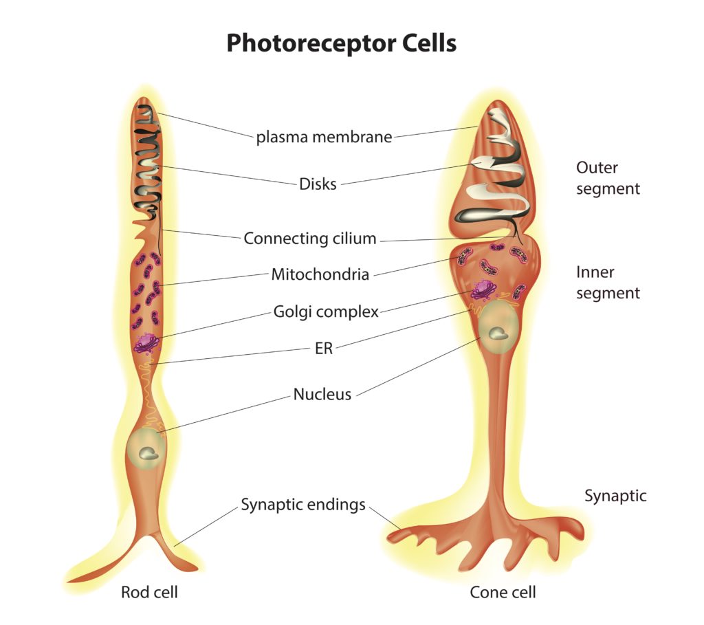
In the rods, which are one of the two types of Photoreceptor cells found in the Retina of the Eye, the detection and conversion of light into a neural signal—a process known as phototransduction—occur within specialized structures. These structures include flat membrane disks and the light-sensitive pigment rhodopsin. Here’s a detailed look at both:
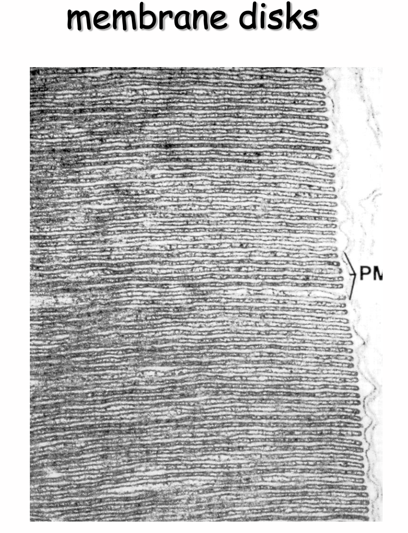
Flat Membrane Disks in Rods
- Structure: Rod cells contain a specialized compartment called the outer segment, which is packed with stacks of flat membrane disks. These disks are not continuous with the cell membrane but are enclosed within it, creating a distinct environment for phototransduction. In rods, the disks are more numerous and densely packed than in cones, which contributes to their high sensitivity to light.
- Function: The primary function of these disks is to house the visual pigment molecules, including rhodopsin, in an organized manner that maximizes the cell’s ability to capture photons of light. The large surface area provided by the disks ensures that a single rod cell can contain millions of rhodopsin molecules, significantly enhancing its light-absorbing capacity.
Rhodopsin in Rods
- Composition: Rhodopsin is a visual pigment found in the membrane disks of rod cells. It is a complex molecule composed of the protein opsin and a light-sensitive chromophore called retinal (specifically, 11-cis-retinal).
- Light Absorption: Rhodopsin is responsible for the initial step in the detection of light. When a photon of light hits rhodopsin, the retinal component absorbs the photon and undergoes a conformational change from the 11-cis form to the all-trans form. This change triggers a series of biochemical reactions within the rod cell.
- Activation of Signal Transduction Pathway: The conformational change in retinal activates the opsin, which in turn activates a G-protein called transducin. Activated transducin triggers a cascade that leads to the reduction of cyclic guanosine monophosphate (cGMP) levels in the cell. As cGMP levels fall, cyclic nucleotide-gated (CNG) ion channels close, leading to the hyperpolarization of the rod cell and a decrease in the release of neurotransmitters to bipolar cells. This change in neurotransmitter release alters the signal sent to the brain, ultimately resulting in the perception of light.
- Regeneration: After activation, rhodopsin must be regenerated to its original state to respond to new light stimuli. This involves several steps, including the conversion of all-trans-retinal back to 11-cis-retinal, which then recombines with opsin to form functional rhodopsin again.
The efficiency and sensitivity of rods in low light conditions are largely due to the abundance of flat membrane disks packed with rhodopsin. This arrangement allows rods to be incredibly sensitive to light, enabling night vision and the perception of movement and shapes in dim conditions.
see also
Tags: neurobiology science
Superlink: 051 ☣Neurobiology 050 🧠Neuroscience
Source
Created: 16-09-24 15:21