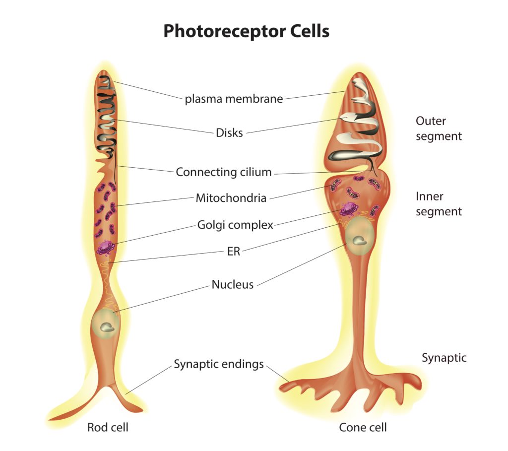
Photoreceptors
chatbot
Photoreceptors are integral components of the visual system, converting light into signals that can be interpreted by the brain to form images. These specialized neurons are located in the retina’s outermost layer and are pivotal in initiating the process of vision.
outer segment: photosensitive part of the cells
Rods:
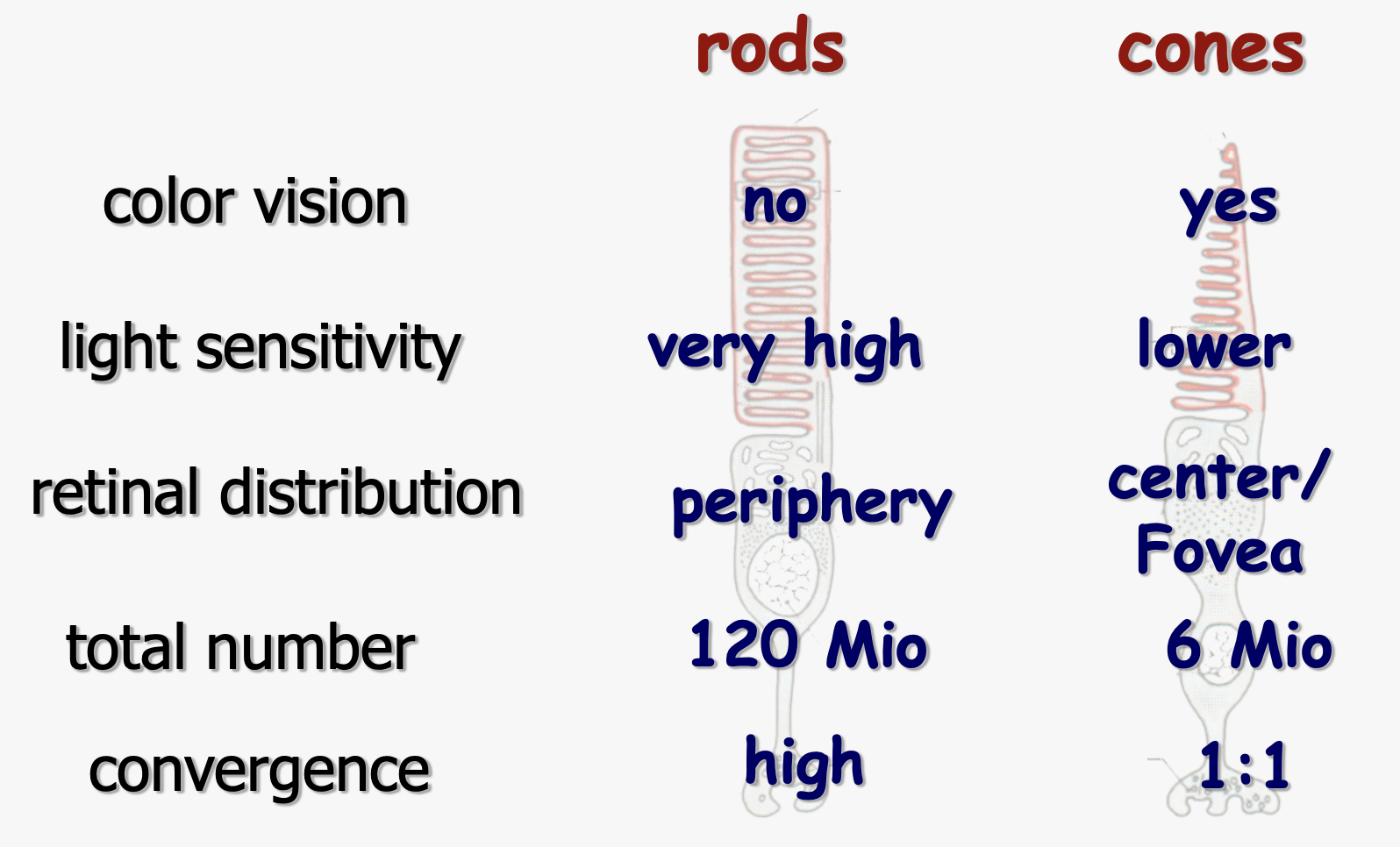
Rods
Rods are designed for low-light conditions, making them essential for night vision and detecting movement in the periphery of our visual field. They are not sensitive to color, contributing mainly to black and white vision.
- With approximately 120 million rods in the human eye, they outnumber cones significantly, highlighting their importance in our ability to see in dim light or at night.
- Rods have a high sensitivity to light but do not provide detailed vision or color differentiation. This is why in low-light conditions, colors are less discernible, and our vision is more blurred.
In the rods, which are one of the two types of Photoreceptor cells found in the Retina of the Eye, the detection and conversion of light into a neural signal—a process known as phototransduction—occur within specialized structures. These structures include flat membrane disks and the light-sensitive pigment rhodopsin. Here’s a detailed look at both:
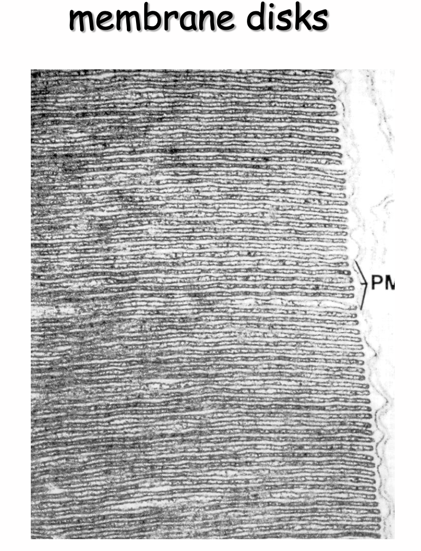
Flat Membrane Disks in Rods
- Structure: Rod cells contain a specialized compartment called the outer segment, which is packed with stacks of flat membrane disks. These disks are not continuous with the cell membrane but are enclosed within it, creating a distinct environment for phototransduction. In rods, the disks are more numerous and densely packed than in cones, which contributes to their high sensitivity to light.
- Function: The primary function of these disks is to house the visual pigment molecules, including rhodopsin, in an organized manner that maximizes the cell’s ability to capture photons of light. The large surface area provided by the disks ensures that a single rod cell can contain millions of rhodopsin molecules, significantly enhancing its light-absorbing capacity.
Rhodopsin in Rods
- Composition: Rhodopsin is a visual pigment found in the membrane disks of rod cells. It is a complex molecule composed of the protein opsin and a light-sensitive chromophore called retinal (specifically, 11-cis-retinal).
- Light Absorption: Rhodopsin is responsible for the initial step in the detection of light. When a photon of light hits rhodopsin, the retinal component absorbs the photon and undergoes a conformational change from the 11-cis form to the all-trans form. This change triggers a series of biochemical reactions within the rod cell.
- Activation of Signal Transduction Pathway: The conformational change in retinal activates the opsin, which in turn activates a G-protein called transducin. Activated transducin triggers a cascade that leads to the reduction of cyclic guanosine monophosphate (cGMP) levels in the cell. As cGMP levels fall, cyclic nucleotide-gated (CNG) ion channels close, leading to the hyperpolarization of the rod cell and a decrease in the release of neurotransmitters to bipolar cells. This change in neurotransmitter release alters the signal sent to the brain, ultimately resulting in the perception of light.
- Regeneration: After activation, rhodopsin must be regenerated to its original state to respond to new light stimuli. This involves several steps, including the conversion of all-trans-retinal back to 11-cis-retinal, which then recombines with opsin to form functional rhodopsin again.
The efficiency and sensitivity of rods in low light conditions are largely due to the abundance of flat membrane disks packed with rhodopsin. This arrangement allows rods to be incredibly sensitive to light, enabling night vision and the perception of movement and shapes in dim conditions.
Link zum Original
Cones:
Cones
- Cones are the photoreceptors responsible for color vision and the ability to see fine details, playing a crucial role in tasks requiring high visual acuity, such as reading or distinguishing faces.
- They are primarily located in the fovea, a small depression in the retina where visual acuity is highest. This central focus area is densely packed with cones and is critical for tasks that require detailed vision.
- The human eye contains about 6 million cones, categorized into three types based on their sensitivity to different wavelengths of light: short (S), medium (M), and long (L). These correspond to blue, green, and red light, respectively, and their combined activity allows us to perceive a wide spectrum of colors.
- Unlike rods, cones require more light to function effectively, which is why in dark environments, colors seem faded or indistinguishable.
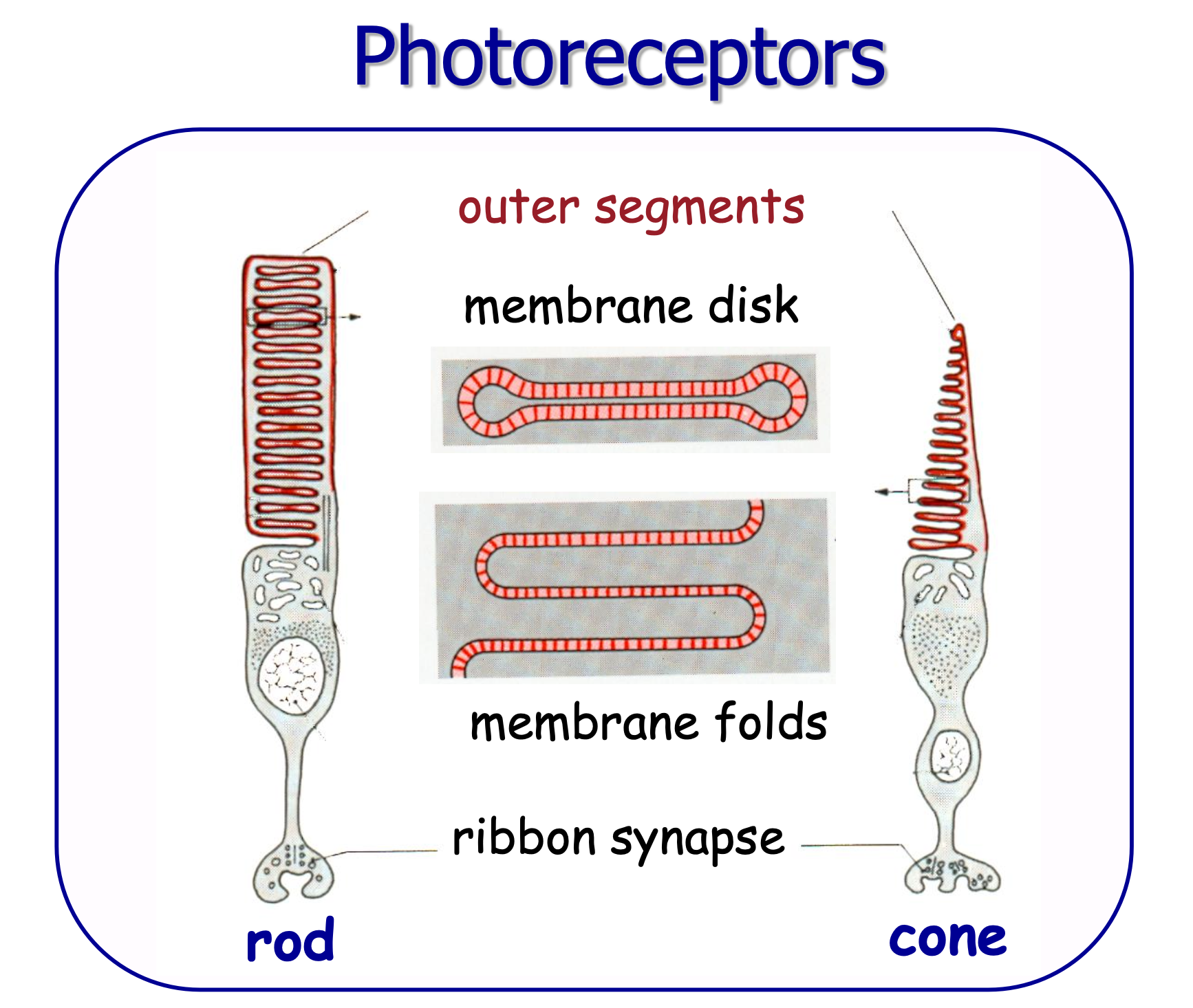
Cones are one of the two types of photoreceptor cells in the retina, responsible for color vision and visual acuity, particularly in well-lit conditions. Unlike rods, which contain stacks of flat membrane disks that are separate from the cell’s plasma membrane, cones feature a pleated structure in their outer segments where the plasma membrane folds back on itself, forming invaginations or infoldings. This structural difference is key to understanding how cones function and how they are adapted to their specific role in the visual system.Pleated Outer Plasma Membrane in Cones
Structure: The outer segment of cone cells is characterized by numerous folds or pleats in the plasma membrane. These folds are continuous with the cell’s outer membrane, unlike the discrete, internalized disks found in rods. This arrangement creates a series of infoldings that increase the surface area available for housing photopigments without detaching from the outer membrane.
Function: The primary function of these pleated membranes is to accommodate the visual pigments necessary for color vision. Conxoes contain different types of photopigments (opsins) that are sensitive to different wavelengths of light, corresponding to blue, green, and red light. The increased surface area provided by the pleated membrane allows for a high concentration of photopigment molecules, which is essential for color discrimination and high spatial acuity.
Phototransduction in Cones: The process of phototransduction in cones is similar to that in Rods, involving the absorption of light by photopigments (in this case, cone opsins), which triggers a biochemical cascade that ultimately changes the electrical charge of the cell and affects neurotransmitter release. However, cones are less sensitive to light than rods and are designed to operate under brighter light conditions. The structural adaptation of the pleated membrane supports this function by optimizing the arrangement of photopigments for the detection of fine detail and color.
Adaptation to Bright Light: The structural differences between rods and cones, including the presence of pleated outer plasma membranes in cones, reflect their adaptation to different lighting conditions. Cones’ pleated membranes allow them to quickly regenerate their photopigments, enabling them to function in bright light and recover quickly from exposure to intense light sources.
In summary, the pleated outer plasma membrane of cones is a specialized adaptation that allows these cells to efficiently capture light and contribute to color vision and visual acuity. This structure supports the high density of photopigments needed for cones’ role in daylight vision, color discrimination, and the perception of fine detail.
Link zum Original


Behaviour of Photoreceptors:
- The process of converting light into an electrical signal is known as phototransduction. This complex biochemical process alters the photoreceptor’s polarity, leading to a change in the release of neurotransmitters and initiating a neural response.
- This response is then refined and processed by other types of neurons within the retina, such as bipolar and ganglion cells, before being relayed to the brain through the optic nerve.
- The brain interprets these signals, allowing us to experience the sensation of vision. The intricate processing of these signals enables us to perceive depth, movement, color, and contrast, forming the rich tapestry of our visual world.
Photoreceptors are fundamental to our understanding and interaction with our environment, translating light into a language the brain can understand and respond to. Their specialized functions, distribution across the retina, and the intricate processes they initiate underscore the complexity and efficiency of the human visual system.

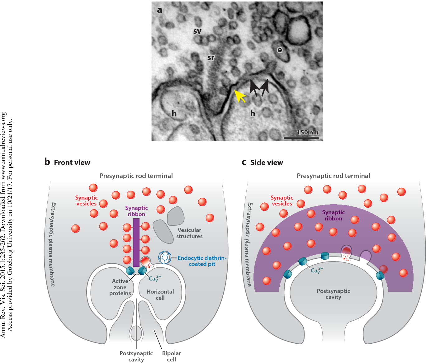
Synaptic ribbon
attracts synaptic vesicles ready for collective exocytosis (“compound fusion”).
chatbot
The synaptic ribbon is a specialized structure found in the synapses of photoreceptor cells (rods and cones) in the retina, as well as in certain other sensory neurons, such as those in the inner ear. This structure plays a crucial role in the continuous and rapid release of neurotransmitters, which is essential for the transmission of visual information from photoreceptors to other neurons in the Retina under a wide range of light conditions.
Structure and Location
- Structure: The synaptic ribbon is an electron-dense, ribbon-like structure anchored to the presynaptic membrane at the base of the photoreceptor cell, where it forms a synaptic connection with the dendrites of bipolar cells and Horizontal Cells. It is typically associated with a large number of synaptic vesicles, which contain neurotransmitters.
- Location: In photoreceptor cells, synaptic ribbons are located in the synaptic terminal, also known as the spherule (in rods) or pedicle (in cones). These terminals are the sites where photoreceptors make synaptic contacts with the secondary neurons of the retina.
Function
- Rapid Neurotransmitter Release: The synaptic ribbon facilitates the rapid and sustained release of neurotransmitters. It does this by organizing a pool of readily releasable synaptic vesicles at the active zone of the synapse, ensuring that neurotransmitter release can keep pace with the constant influx of visual information, especially in varying light conditions.
- Continuous Signaling: Unlike conventional synapses, which may fire in a more “on-off” fashion, the synaptic ribbon enables continuous signaling by ensuring a steady flow of neurotransmitters. This is crucial for the encoding of graded changes in light intensity over a broad dynamic range, characteristic of visual processing.
- Sensitivity to Light Changes: By facilitating the rapid and continuous release of neurotransmitters, synaptic ribbons allow photoreceptor cells to respond swiftly and accurately to changes in light intensity. This contributes to the high sensitivity and temporal resolution of the visual system, enabling it to detect and process visual stimuli with remarkable precision.
Mechanism
The exact mechanism by which synaptic ribbons facilitate Neurotransmitter release is still under investigation, but it is known that they play a key role in tethering and positioning synaptic vesicles near the active zone. This ensures that vesicles can be rapidly released in response to an influx of calcium ions when the photoreceptor is stimulated by light. The continuous and regulated release of neurotransmitters at these ribbon synapses is essential for the transmission of visual signals to the brain, allowing for the perception of light, color, and movement.
In summary, the synaptic ribbon is a critical component of the photoreceptor synapse, enabling the high-fidelity transmission of visual information from the Retina to the brain by facilitating the rapid and sustained release of neurotransmitters. This unique structure is essential for the continuous and precise signalling required for visual processing.
see also
Tags: neurobiology science
Superlink: 051 ☣Neurobiology 050 🧠Neuroscience
034 💪🦵Physiology
Eye
Retina
Source
Created: 16-09-24 13:49