natural sleep or anesthesia are associated with a 60% increase in the interstitial space, resulting in a striking increase in convective exchange of cerebrospinal fluid with interstitial fluid.
convective fluxes of interstitial fluid increased the rate of β-amyloid clearance during sleep.
the restorative function of sleep may be a consequence of the enhanced removal of potentially neurotoxic waste products that accumulate in the awake central nervous system.
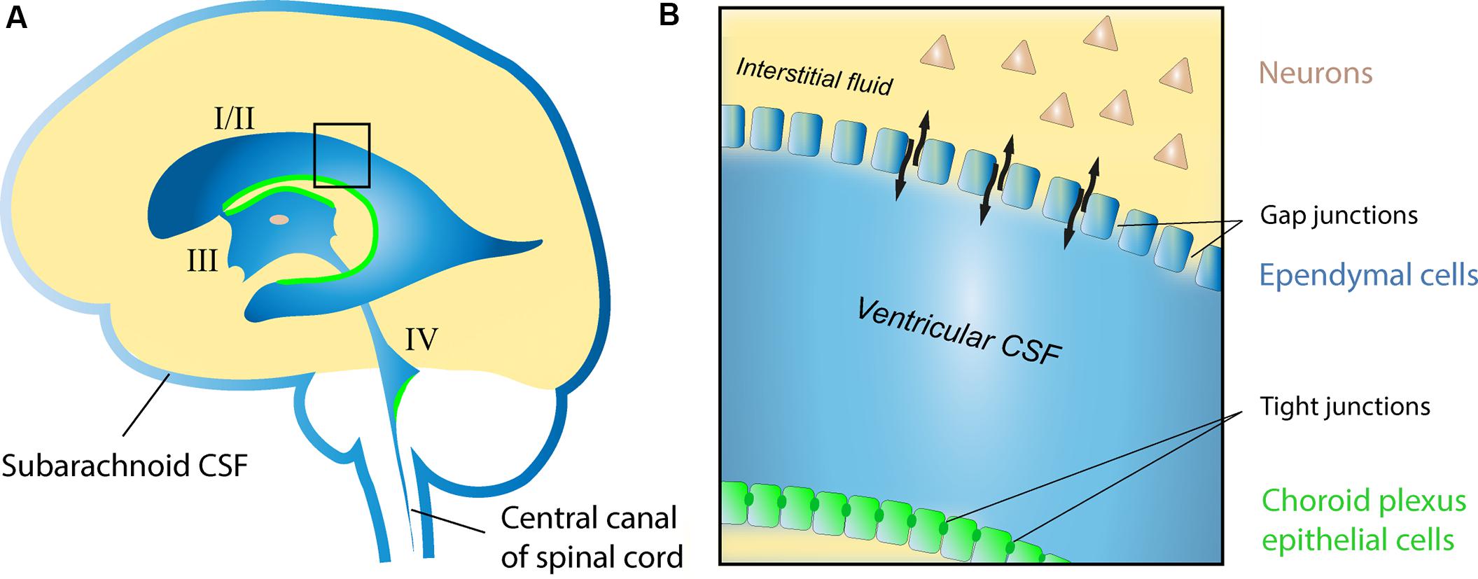
Proteins linked to neurodegenerative diseases, including β-amyloid (Aβ) (8), α-synuclein (9), and tau (10), are present in the interstitial space surrounding cells of the brain.
cerebrospinal fluid (CSF) recirculates through the brain, interchanging with interstitial fluid (ISF) and removing interstitial proteins, including Aβ
cerebrospinal fluid (CSF) recirculates through the brain, interchanging with interstitial fluid (ISF) and removing interstitial proteins, including Aβ
convective movement of ISF is a substantial contributor to the removal of interstitial waste products and other products of cellular activity. The interstitial concentration of Aβ is higher in awake than in sleeping rodents and humans, possibly indicating that wakefulness is associated with increased Aβ production

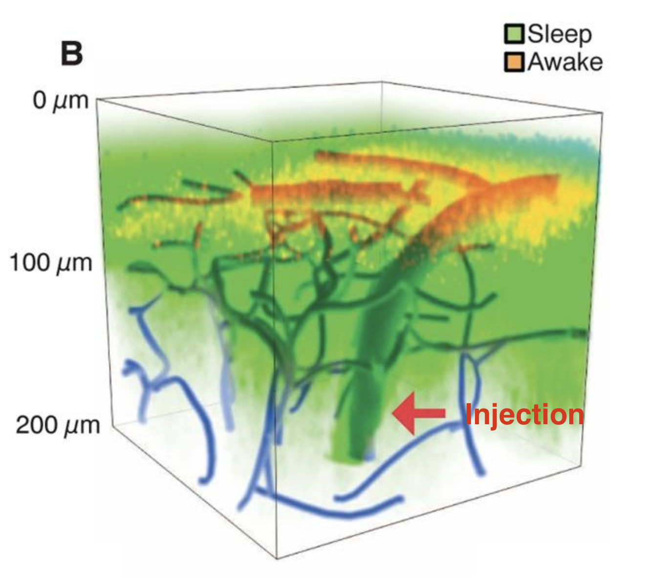
glymphatic CSF influx is sharply suppressed in conscious alert mice as compared with naturally sleeping or anesthetized littermates
the cortical interstitial volume fraction is 13-15% in the awake state as compared to 22 to 24% in sleeping or anesthetized mice.
This effect of arousal state on interstitial volume likely holds major implications for diffusion of neurotransmitters, such as glutamate
65% of exogenously delivered Aβ is cleared by the glymphatic system
Aβ clearance did not differ between sleeping and anesthetized mice. Because Aβ is also removed from CNS via receptor-mediated transport across the blood-brain barrier (24), we also analyzed the clearance of an inert tracer, 14C-inulin.14C- inulin was cleared more efficiently (greater than twofold) in sleeping and anaesthetized mice as compared with awake
The observation that anesthesia increases glymphatic influx and efflux (Figs. 1 and 3), suggests that it is not circadian rhythm but rather the sleep-wake state itself that determines the volume of the interstitial space and therefore the efficiency of glymphatic solute clearance.
noradrenaline regulates the activity of membrane transporters and channels that control cell volume
adrenergic signaling plays an important role in modulating not only cortical neuronal activity but also the volume of the interstitial space.
NE triggers rapid changes in neural activity (27, 28), which in turn can modulate the volume of the interstitial space volume (29). Nevertheless, additional analysis is clearly required to determine which cell types contribute to expansion of the interstitial space volume during sleep, anesthesia, or blockade of NE receptors (Figs. 2, B to D, and 4D).
Because of the high sensitivity of neural cells to their environment, it is essential that waste products of neural metabolism are quickly and efficiently removed from the brain interstitial space.
An extension of the findings reported here is that the restorative function of sleep may be due to the switching of the brain into a functional state that facilitates the clearance of degradation products of neural activity that accumulate during wakefulness.
suggestions
possibility is that the awake brain
state is linked to a reduction in the volume of the interstitial space because a constricted interstitial space would increase resistance to convective fluid movement and suppress CSF influx.
We hypothesized that adrenergic signaling in the awake state modifies cell volume and thus the size of the interstitial space.
Taken altogether available data indicate that sleep increases perivascular and non-perivascular clearances for amyloid-β which reduces its concentration and may have long-term consequences for the formation of plaques and cerebral arterial deposits.
nice to know, aber kommt nicht rein
Influx of CSF is in part driven by arterial pulse waves that propel the movement of CSF inward along periarterial spaces
vielleicht:
anesthesia consistently increased the interstitial space volume fraction by >60%
from other sources
We show that Aβ accumulation preferentially starts in the precuneus, medial orbitofrontal, and posterior cingulate cortices, i.e., several of the core regions of the The Default Mode Network and Mind Wandering
Source:
https://www.nature.com/articles/s41467-017-01150-x
Palmqvist, S., Schöll, M., Strandberg, O., Mattsson, N., Stomrud, E., Zetterberg, H., Blennow, K., Landau, S., Jagust, W., & Hansson, O. (2017). Earliest accumulation of β-amyloid occurs within the default-mode network and concurrently affects brain connectivity. Nature Communications, 8(1), 1-13. https://doi.org/10.1038/s41467-017-01150-x
Frage:
Was bedeutet das?
Dass man nicht mehr so gut Mind Wandering betreiben kann?
Kommt man dann nicht mehr auf neue und gute Ideen?
Sleep Deprivation
Sleep deprivation reduces learning, impairs performance in cognitive tests, prolongs reaction time, and is a common cause of seizures (3, 4). In the most extreme case, continuous sleep deprivation kills rodents and flies within a period of days to weeks (5, 6).
see also
Tags: neuroscience science
Superlink: 050 🧠Neuroscience
051 ☣Neurobiology
Memory in Sleep
Sleep Drives Metabolite Clearance from the Adult Brain
Source
Xie, L., Kang, H., Xu, Q., Chen, M. J., Liao, Y., Thiyagarajan, M., Christensen, D. J., Nicholson, C., Iliff, J. J., Takano, T., Deane, R., & Nedergaard, M. (2013). Sleep Drives Metabolite Clearance from the Adult Brain. Science (New York, N.Y.), 342(6156). https://www.ncbi.nlm.nih.gov/pmc/articles/PMC3880190/
Created: 30-01-24 16:41