Cones
- Cones are the photoreceptors responsible for color vision and the ability to see fine details, playing a crucial role in tasks requiring high visual acuity, such as reading or distinguishing faces.
- They are primarily located in the fovea, a small depression in the retina where visual acuity is highest. This central focus area is densely packed with cones and is critical for tasks that require detailed vision.
- The human eye contains about 6 million cones, categorized into three types based on their sensitivity to different wavelengths of light: short (S), medium (M), and long (L). These correspond to blue, green, and red light, respectively, and their combined activity allows us to perceive a wide spectrum of colors.
- Unlike rods, cones require more light to function effectively, which is why in dark environments, colors seem faded or indistinguishable.
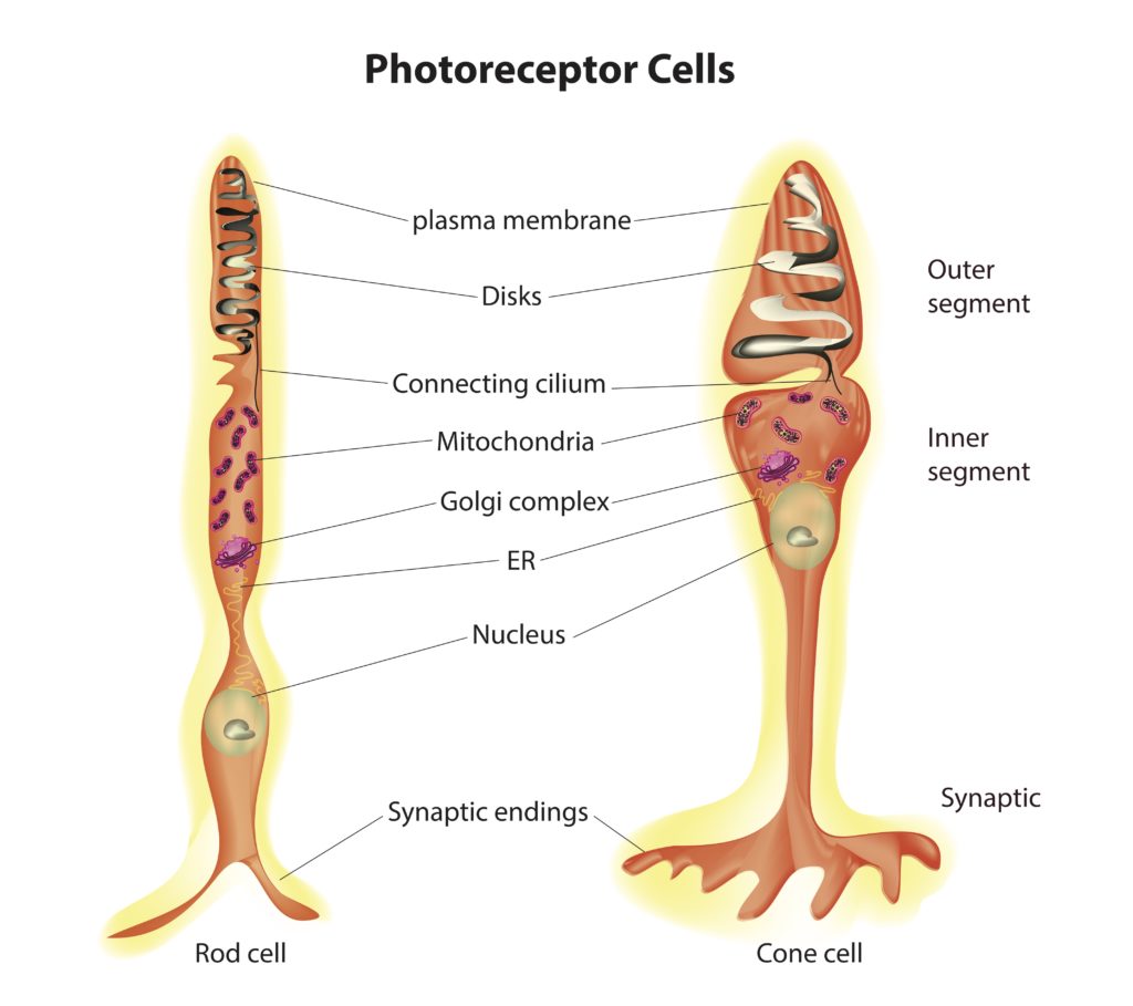
Cones are one of the two types of photoreceptor cells in the retina, responsible for color vision and visual acuity, particularly in well-lit conditions. Unlike rods, which contain stacks of flat membrane disks that are separate from the cell’s plasma membrane, cones feature a pleated structure in their outer segments where the plasma membrane folds back on itself, forming invaginations or infoldings. This structural difference is key to understanding how cones function and how they are adapted to their specific role in the visual system.
Pleated Outer Plasma Membrane in Cones
-
Structure: The outer segment of cone cells is characterized by numerous folds or pleats in the plasma membrane. These folds are continuous with the cell’s outer membrane, unlike the discrete, internalized disks found in rods. This arrangement creates a series of infoldings that increase the surface area available for housing photopigments without detaching from the outer membrane.

-
Function: The primary function of these pleated membranes is to accommodate the visual pigments necessary for color vision. Conxoes contain different types of photopigments (opsins) that are sensitive to different wavelengths of light, corresponding to blue, green, and red light. The increased surface area provided by the pleated membrane allows for a high concentration of photopigment molecules, which is essential for color discrimination and high spatial acuity.
-
Phototransduction in Cones: The process of phototransduction in cones is similar to that in Rods, involving the absorption of light by photopigments (in this case, cone opsins), which triggers a biochemical cascade that ultimately changes the electrical charge of the cell and affects neurotransmitter release. However, cones are less sensitive to light than rods and are designed to operate under brighter light conditions. The structural adaptation of the pleated membrane supports this function by optimizing the arrangement of photopigments for the detection of fine detail and color.
-
Adaptation to Bright Light: The structural differences between rods and cones, including the presence of pleated outer plasma membranes in cones, reflect their adaptation to different lighting conditions. Cones’ pleated membranes allow them to quickly regenerate their photopigments, enabling them to function in bright light and recover quickly from exposure to intense light sources.
In summary, the pleated outer plasma membrane of cones is a specialized adaptation that allows these cells to efficiently capture light and contribute to color vision and visual acuity. This structure supports the high density of photopigments needed for cones’ role in daylight vision, color discrimination, and the perception of fine detail.
see also
Tags: neurobiology science
Superlink: 051 ☣Neurobiology 050 🧠Neuroscience
034 💪🦵Physiology
Vision
Retina
Eye
Photoreceptor cells
Source
Created: 16-09-24 15:38