Dark current
chatbot
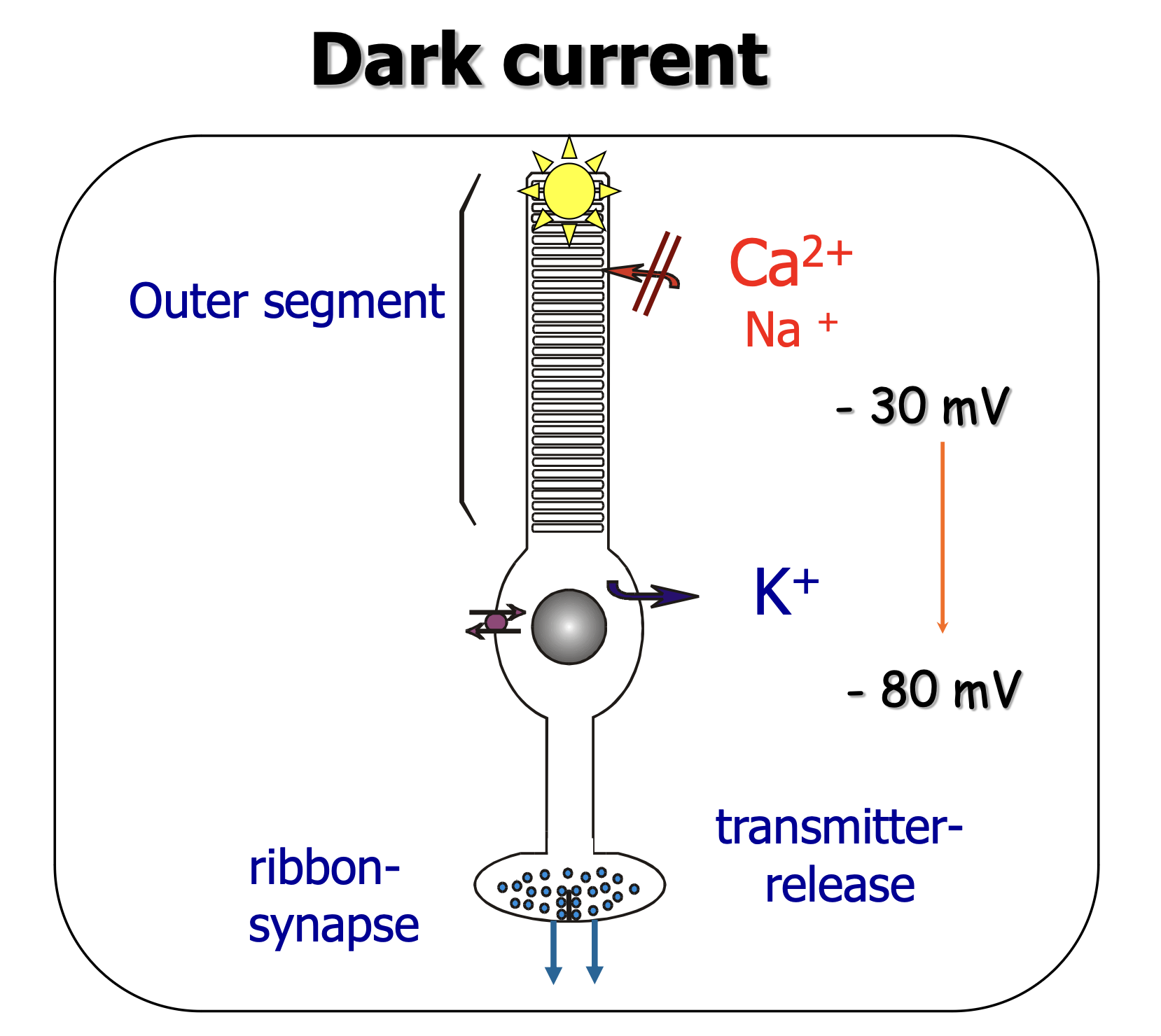
The “dark current” in the retina refers to a continuous flow of ions that occurs in the photoreceptor cells (rods and cones) in the absence of light. This physiological process is crucial for the photoreceptors’ ability to respond to changes in illumination and contribute to vision. Here’s a more detailed explanation of how it works:
Mechanism of Dark Current
- In the Dark: In the absence of light, photoreceptor cells exhibit a unique activity known as the dark current. During this state, the Photoreceptor cells are depolarized, meaning there is a relatively positive charge inside the cell compared to the outside. This depolarization is maintained by the continuous influx of sodium ions (Na+) into the cell through specialized channels in the cell membrane called cyclic nucleotide-gated (CNG) channels.
- Role of cGMP: The opening of these CNG channels is regulated by the concentration of cyclic guanosine monophosphate (cGMP) within the cell. In the dark, levels of cGMP are high, keeping the CNG channels open and allowing the influx of Na+ ions, which generates the dark current.
- Energy Consumption: Maintaining the dark current requires significant metabolic energy because the photoreceptor cells must continuously pump the incoming sodium ions back out to maintain the electrochemical gradient. This process is mediated by the Na+/K+ ATPase pump.
The difference of cGMP and cAMP
cAMP and cGMP
They are very similar in structure, but have very different functions.
Response to Light
- Light Exposure: When photoreceptor cells are exposed to light, the pigment molecule rhodopsin (in rods) or photopsins (in cones) undergoes a conformational change. This change activates a G-protein called transducin, leading to a cascade of intracellular events that result in the reduction of cGMP levels.
- Closure of CNG Channels: The decrease in cGMP causes the CNG channels to close, stopping the influx of sodium ions and leading to the hyperpolarization of the photoreceptor cell (making the inside of the cell more negative). ⇒ Action Potential
- Signal Transmission: This change in the electrical state of the photoreceptor cell reduces the release of neurotransmitters at the synapse with bipolar cells, altering the signal that is eventually transmitted to the brain via the retinal ganglion cells.
Phototransduction
photoreceptors in the dark
In the dark
- photoreceptors are depolarized in the darkness
- they release the Neurotransmitter Glutamate
- on-bipolar cells possess inhibitory glutamate receptors, i.e. they become hyperpolarized
- connected retinal ganglion cells reduce their firing frequency („off- response“)
what also happens:
3. • off-bipolar cells possess excitatory glutamate receptors, i.e. they become depolarized by glutamate
4. connected retinal ganglion cells increase their firing frequency („on- response“)back in the light
- photoreceptors are hyperpolarized in the light
- they no longer release the neurotransmitter glutamate
- on-bipolar cells return to a depolarized state
- connected retinal ganglion cells increase their firing frequency („on- response“)
what also happens:
3. off-bipolar cells return to a hyperpolarized state
4. connected retinal ganglion cells decrease their firing frequency („off- response“)Phototransduction, the process by which light is converted into electrical signals in the retina, exhibits distinct differences between light and dark conditions. These differences are fundamental to how visual information is processed and transmitted to the brain.
In the Dark:
- Photoreceptor Depolarization: In the absence of light, photoreceptor cells (rods and cones) are depolarized due to the “dark current.” This state is maintained by the continuous influx of sodium and calcium ions through cyclic nucleotide-gated (CNG) channels, which are kept open by high levels of cyclic guanosine monophosphate (cGMP).
- Neurotransmitter Release: The depolarized state of photoreceptors leads to the constant release of the neurotransmitter glutamate.
- Bipolar Cell Response: Glutamate has different effects on bipolar cells due to the types of glutamate receptors they possess. On-bipolar cells, which have inhibitory glutamate receptors, become hyperpolarized and less active. Off-bipolar cells, on the other hand, have excitatory glutamate receptors and become depolarized, leading to increased activity.
- Retinal Ganglion Cell Activity: The activity of connected retinal ganglion cells is modulated accordingly; those connected to on-bipolar cells reduce their firing frequency (“off-response”), while those connected to off-bipolar cells increase their firing frequency (“on-response”).
In the Light:
- Photoreceptor Hyperpolarization: Exposure to light induces a conformational change in the photopigment molecules (rhodopsin in rods, photopsins in cones), leading to a cascade of events that culminates in the reduction of cGMP levels. This causes CNG channels to close, stopping the influx of ions and leading to the hyperpolarization of the photoreceptor cells.
- Reduction in Neurotransmitter Release: The hyperpolarization of photoreceptors in the light significantly reduces or stops the release of glutamate.
- Bipolar Cell Response Reversal: The reduction in glutamate release leads to a reversal in the response of bipolar cells. On-bipolar cells, no longer inhibited by glutamate, return to a depolarized state and become more active. Off-bipolar cells, lacking excitatory glutamate input, return to a hyperpolarized state and become less active.
- Retinal Ganglion Cell Activity Adjustment: The activity of retinal ganglion cells is adjusted once more; those connected to on-bipolar cells increase their firing frequency (“on-response”), while those connected to off-bipolar cells decrease their firing frequency (“off-response”).
Summary:
The key difference in phototransduction between light and dark conditions lies in the state of photoreceptor polarization and the subsequent effects on neurotransmitter release and bipolar cell activity. In the dark, photoreceptors are depolarized and release glutamate, leading to specific responses in bipolar and retinal ganglion cells. In the light, photoreceptors are hyperpolarized, reducing glutamate release and reversing the responses of bipolar and retinal ganglion cells. This dynamic interplay allows the visual system to accurately process and transmit information about the visual environment to the brain.
Link zum Original
summary of dark current
The dark current is thus a fundamental aspect of the phototransduction process, enabling photoreceptor cells to rapidly respond to changes in light intensity and contribute to the formation of visual images.
see also
Tags: neurobiology science
Superlink: 051 ☣Neurobiology 050 🧠Neuroscience
034 💪🦵Physiology
Rods
Cones
Photoreceptor cells
Source
Created: 16-09-24 15:04