Hippocampus
General
The Hippocampus represents a fundamental neurobiological structure for learning and memory, cognitive and emotional functions. The hippocampus is a brain region with high neuronal network activity and Plasticity, that is highly vulnerable to various disruptive events such as hypoxia/ischemia, seizures, head trauma, chronic severe stress, and degenerative processes. This vulnerability is associated with excitatory neurotransmitter overstimulation, high intracellular calcium and long-lasting calpain activation, disruption of energetic metabolism, increased production of reactive oxygen species and high stress-related glucocorticoid level (Liang et al., 2016).
Morphology of the hippocampus**
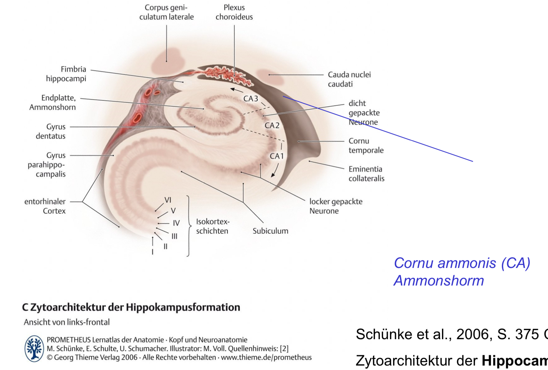
The hippocampus represents a temporal brain structure that belongs to the limbic lobe. In adult humans, it can reach 5 cm in length and 1 cm in width and is formed by two allocortex laminae: the cornu ammonis (CA) and the dentate gyrus. The first mentioned structure can be separated into four arched areas, CA1, CA2, CA3, and CA4, depending on the cellular morphology of the neurons (Fig. 4). The dentate gyrus is a U-shaped structure, found in contact especially with the CA4 region, which fits into the dent of the gyrus. The hippocampus can be divided also into three major parts, the head, the body, and the tail, which represent the narrowest posterior part of the arc. The major hippocampal efferent pathways form the fimbria, which separates progressively from the tail of the hippocampus and thus reveals the gyrus fasciolaris, which is composed of CA3 neurons. The tail of the hippocampus has medial contact with the fimbria and constitutes the floor of the ventricular atrium. The body of the hippocampus is part of the temporal horn’s floor of the lateral ventricle and is bordered by the fimbria in the medial part and by the ventricular protrusion of the collateral sulcus in the lateral part. The hippocampal body holds also an extraventricular side, which is isolated from the subiculum through the hippocampal sulcus. The dilated anterior segment of the hippocampus represents the head (Duvernoy, Cattin, & Risold, 2013; Tatu & Vuillier, 2014).
Regions
chatbot
The regions CA1, CA2, CA3, and CA4 are subdivisions of the hippocampus, specifically within the cornu ammonis, which is crucial for various brain functions including learning and memory. Each of these regions has unique characteristics and plays specific roles in the hippocampal circuitry:
CA1
- CA1: This region is the first segment in the hippocampal circuitry that receives input from the CA3 area through the Schaffer collaterals and sends outputs to both the subiculum and entorhinal cortex, making it a key relay in the flow of information. CA1 is particularly important for the consolidation of long-term memory and is highly susceptible to damage from hypoxia and ischemia.
not chatbot: - The results of our study indicate that hippocampal CA1 neurons play a role in acquisition of the association between auditory cue and direction choice. However, there is no evidence yet that the present memory task depends on the hippocampus and that memory retrieval becomes independent from the hippocampus.
CA2
- CA2: CA2 is a small but distinct region situated between CA1 and CA3, often characterized by its resistance to certain types of damage that affect CA1 and CA3, such as damage from epilepsy. It has unique connectivity and is involved in social memory processing, among other functions. The CA2 region has robust connections with both the CA1 and CA3 regions, as well as with the entorhinal cortex, suggesting its role as an intermediary in hippocampal processing.
CA3
- CA3: CA3 receives inputs from the dentate gyrus and is known for its extensive recurrent collaterals, which allow for a high degree of intraregional connectivity. This region plays a critical role in the initial formation and retrieval of spatial memory and associative memory. CA3’s unique architecture is thought to support pattern completion, a process essential for memory recall.
CA4
- CA4: This region is embedded within the dentate gyrus and acts as a transition zone between the dentate gyrus and CA3. CA4 neurons are influenced by inputs from the dentate gyrus and project to CA3, facilitating the flow of information from the dentate gyrus to CA3. This area is involved in the preprocessing of sensory inputs before they are relayed to CA3 for further processing and memory encoding.
Each of these regions contributes to the complex functions of the hippocampus, highlighting the importance of their distinct but interconnected roles in neuronal network activity, plasticity, and the brain’s overall capacity for learning and memory.
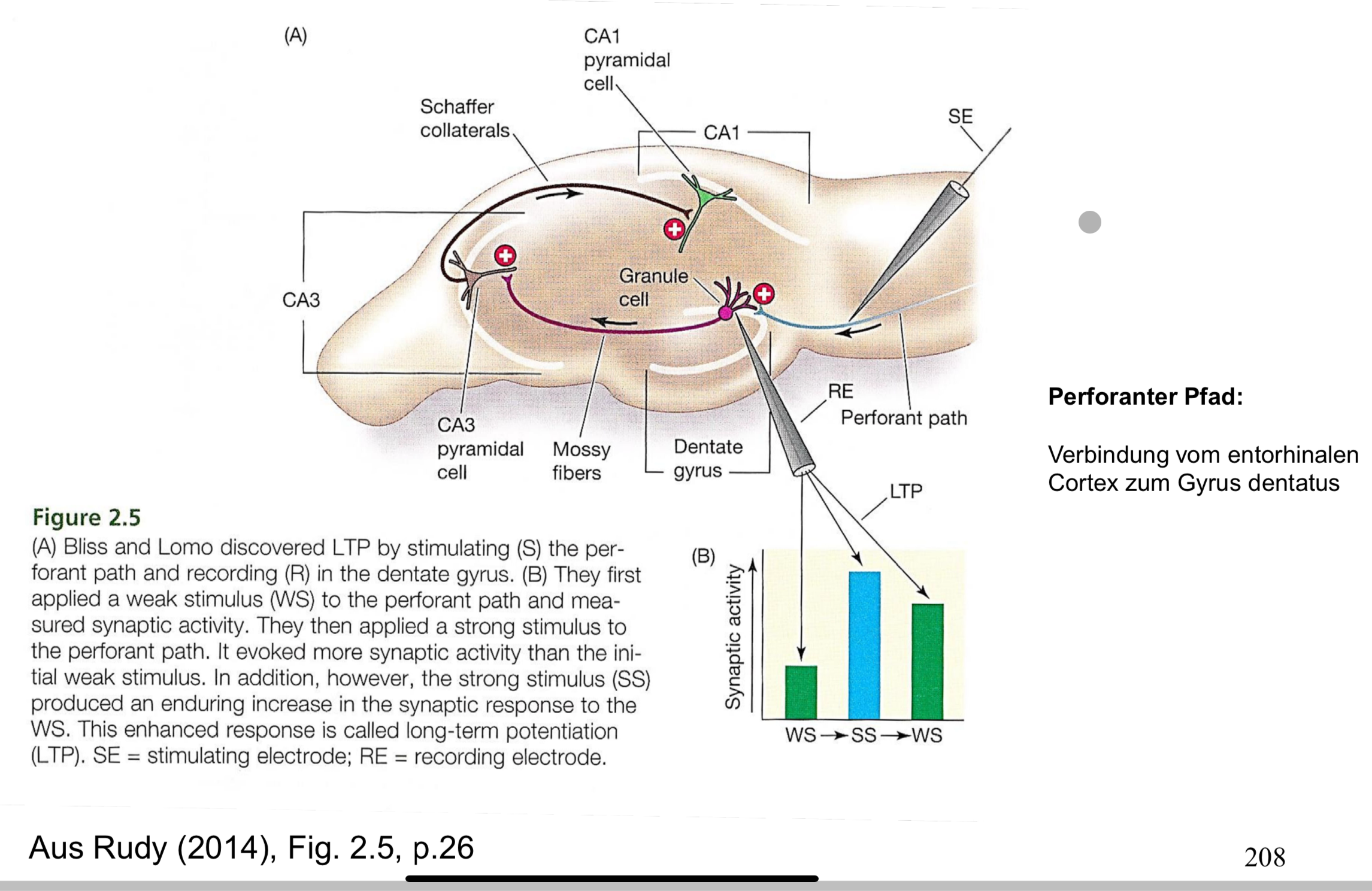
The three synapses of the hippocampus where LTP occurs (+):
- Stimulation of the perforant path from the entorhinal cortex to neurons in the dentate gyrus (initially used by Lomo & Bliss)
- Stimulation of the mossy fibers, reaching CA3 pyramidal cells
- Stimulation of the Schaffer collaterals in the CA3 region, reaching CA1 pyramidal cells
Entorhinal Cortex (via perforant path) → Dentate gyrus (via mossy fibers) → CA3 pyramidal cells (via Schaffer collaterals) → CA1 pyramidal cells
Perforant path
chatbot
The perforant path is a crucial neural pathway in the hippocampus, an area of the brain integral to memory formation and spatial navigation. Originating in the Entorhinal Cortex, the perforant path is the primary conduit of cortical input into the hippocampus, specifically connecting to the dentate gyrus and the CA3 region of the hippocampus. This pathway plays a pivotal role in the encoding, consolidation, and retrieval of memories. The entorhinal cortex, where this pathway begins, is a critical interface between the hippocampus and the neocortex, involved in processing sensory and spatial information before it is transmitted to the hippocampus. The integrity of the perforant path is essential for cognitive function, and damage to this pathway has been implicated in neurodegenerative diseases such as Alzheimer’s disease, where it is one of the first regions to show pathological changes.
see also
Tags: gehirnregion
Superlink: 050 🧠Neuroscience
Hippocampal Memory Consolidation During Sleep A Comparison of Mammals and Birds - PMC
LTP - long term potentiation
LTD - Long-Term Depression
Quellen
Erstellt: 05-10-21 17:04