Retina
- Retina is part of the Central Nervous System (CNS), forming from the diencephalon during embryonic development.
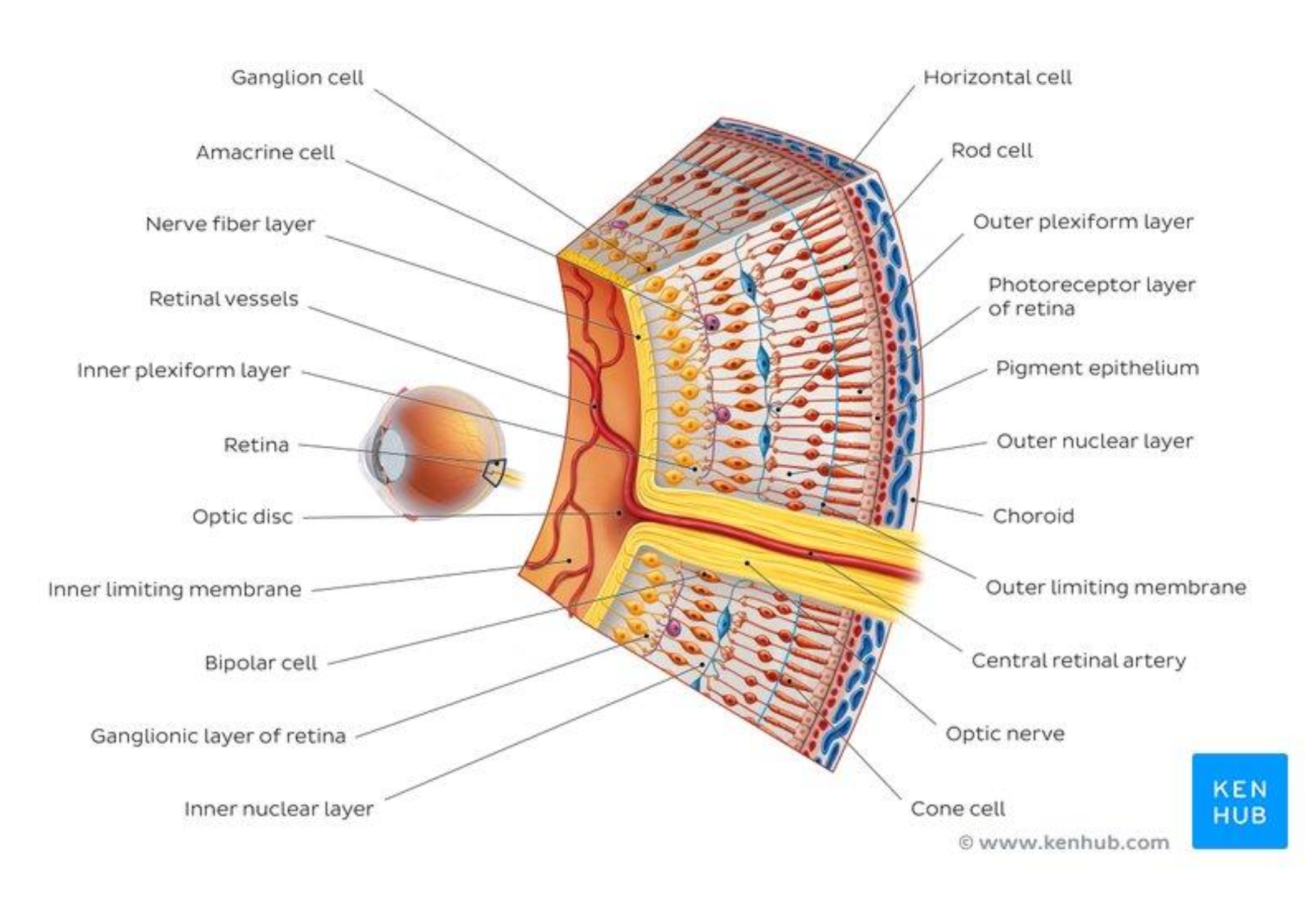
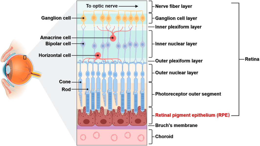
- Contains a limited number of cell types in a highly ordered network.
- Outermost layer contains Photoreceptor cells (rods and cones) surrounded by pigment epithelium for support and light shielding.
- Visual signals from photoreceptors transfer radially to bipolar cells, then to retinal ganglion cells.
- Axons of retinal ganglion cells form the optic nerve, exporting signals to the brain.
- Blind spot: Area where the optic nerve leaves, containing no photoreceptors.
- Two horizontal paths of information processing: Horizontal Cells (between photoreceptors) and amacrine cells (between bipolar and retinal ganglion cells).
- Photoreceptor cells: Rods (dim light vision) and Cones (color vision under good illumination).
- Phototransduction: Absorption of light and conversion into a neural signal.
- Outer segment of photoreceptors: Rods have flat membrane disks with rhodopsin; Cones have a pleated outer plasma membrane.
- Synaptic ribbon in photoreceptors facilitates collective exocytosis.
- Distribution: Cones concentrated in the fovea (high visual acuity); Rods predominate in the peripheral region.
- Connectivity: Cones in fovea connect to retinal ganglion cells in a 1:1 relationship; Rods have over 1000 cells synapsing onto a single retinal ganglion cell, enhancing light sensitivity.
see also
Tags: neurobiology science
Superlink: 051 ☣Neurobiology 050 🧠Neuroscience
034 💪🦵Physiology
Source
Created: 16-09-24 14:05